| 结构式 | 名称/CAS号 | 全部文献 |
|---|---|---|
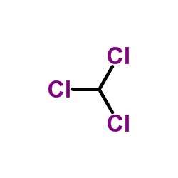 |
氯仿
CAS:67-66-3 |
|
 |
六甲基二硅氮烷
CAS:999-97-3 |
|
 |
N,N-二甲基甲酰胺
CAS:68-12-2 |
|
 |
锇酸酐
CAS:20816-12-0 |
|
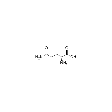 |
L-谷氨酰胺
CAS:56-85-9 |
|
 |
活性炭
CAS:7440-44-0 |
|
 |
炭黑
CAS:1333-86-4 |
|
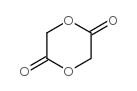 |
乙交酯
CAS:502-97-6 |
|
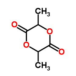 |
DL-丙交酯
CAS:95-96-5 |
|
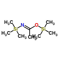 |
N,O-双(三甲基硅基)乙酰胺
CAS:10416-59-8 |