Crystal structure of the complex formed between a group I phospholipase A2 and a naturally occurring fatty acid at 2.7 A resolution.
Garima Singh, Jayasankar Jasti, K Saravanan, Sujata Sharma, Punit Kaur, A Srinivasan, Tej P Singh
文献索引:Protein Sci. 14(2) , 395-400, (2005)
全文:HTML全文
摘要
This is the first evidence of a naturally bound fatty acid to a group I Phospholipase A(2) (PLA(2)) and also to a PLA(2) with Asp 49. The fatty acid identified as n-tridecanoic acid is observed at the substrate recognition site of PLA(2) hydrophobic channel. The complex was isolated from the venom of Bungarus caeruleus (Common Indian Krait). The primary sequence of the PLA(2) was determined using the cDNA method. Three-dimensional structure has been solved by the molecular replacement method and refined using the CNS package to a final R factor of 19.8% for the data in the resolution range from 20.0 to 2.7 A. The final refined model is comprised of 912 protein atoms, one sodium ion, one molecule of n-tridecanoic acid, and 60 water molecules. The sodium ion is located in the calcium-binding loop with a sevenfold coordination. A characteristic extra electron density was observed in the hydrophobic channel of the enzyme, into which a molecule of n-tridecanoic acid was clearly fitted. The MALDI-TOF measurements of the crystals had earlier indicated an increase in the molecular mass of PLA(2) by 212 Da over the native PLA(2). A major part of the ligand fits well in the binding pocket and interacts directly with His 48 and Asp 49. Although the overall structure of PLA(2) in the present complex is similar to the native structure reported earlier, it differs significantly in the folding of its calcium-binding loop.
相关化合物
| 结构式 | 名称/CAS号 | 分子式 | 全部文献 |
|---|---|---|---|
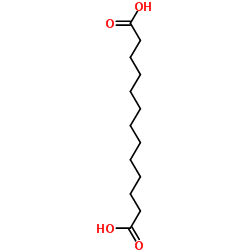 |
十三碳二元酸
CAS:505-52-2 |
C13H24O4 |
|
Pseudohomogeneous kinetic study on a two-liquid-phase fermen...
1994-01-01 [Chin. J. Biotechnol. 10(4) , 271-82, (1994)] |
|
[Search for yeast producers of brassylic and sebacic fatty a...
2004-01-01 [Prikl. Biokhim. Mikrobiol. 40(5) , 533-5, (2004)] |
|
[Studies on microbial production of undecane 1, 11-dicarboxy...
1999-06-01 [Wei Sheng Wu Xue Bao 39(3) , 279-81, (1999)] |
|
Macrocyclic musk compounds--an absence of genotoxicity in th...
2002-09-05 [Toxicol. Lett. 135(1-2) , 155-63, (2002)] |
|
Mass spectrometric identification of 2-hydroxy-sebacic acid ...
1986-06-01 [Biomed. Environ. Mass Spectrom. 13(6) , 315-8, (1986)] |