| Structure | Name/CAS No. | Articles |
|---|---|---|
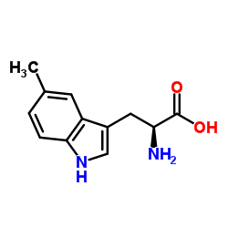 |
5-Methyltryptophan
CAS:951-55-3 |
|
 |
Indole-3-acetamide
CAS:879-37-8 |