| Description |
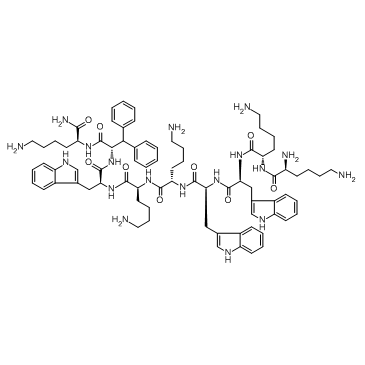
LTX-315 is an oncolytic peptide with potent anticancer activity; inhibits MRC-5, A20 and AT84 with IC50s of 34.3, 8.3 and 11 µM, respectively.
|
| Related Catalog |
|
| Target |
IC50: 34.3 µM (MRC-5), 8.3 µM (A20), 11 µM (AT84)[1]
|
| In Vitro |
LTX-315 is found to be equipotent against drug-resistant cancer cells, nontoxic towards red blood cells, shows high plasma protein binding and is quite rapidly degraded to non-toxic metabolites. LTX-315 induces rapid killing of cancer cells. The oncolytic activity of LTX-315 stems from both a direct lytic effect on the plasma membrane in addition to permeabilization of the mitochondrial membrane, leading to cellular death by necrosis and release of tumor antigens. Treatment of cancer cells with LTX-315 causes the release of several danger signals (DAMPs) that are associated with immunogenic cell death and stimulation of adaptive immune responses[1].
|
| In Vivo |
Intratumoral administration of LTX-315 has resulted in complete regression and systemic tumor specific immune responses in several preclinical models[1]. Intratumoral administration of LTX-315 resulted in tumor necrosis and the infiltration of immune cells into the tumor parenchyma followed by complete regression of the tumor in the majority of the animals. LTX-315 induced the release of danger-associated molecular pattern molecules such as the high mobility group box-1 protein in vitro and the subsequent upregulation of proinflammatory cytokines such as interleukin (IL) 1β, IL6 and IL18 in vivo. Animals cured by LTX-315 treatment are protected against a re-challenge with live B16 tumor cells both intradermally and intravenously[2].
|
| Cell Assay |
Tumor cells are incubated for 4 hours with 10 concentrations of LTX-315 in 1/4 dilution step with a top dose of 400 µM, with 1% (final concentration) Triton X-100 as positive control and FBS-free culture medium as negative control. Cell cytotoxicity is measured using the MTS assay[1].
|
| Animal Admin |
Mice: Tumor cells are harvested, ished in RPMI-1640 and injected intradermally (i.d.) into the right side of the abdomen in C57BL/6 mice. Palpable tumors are injected i.t. with single doses of LTX-315 or LTX-328 dissolved in saline (1.0 mg peptide/50 μL saline) once a day for 3 consecutive days, and the vehicle control is saline only (0.9% NaCl in sterile water). Tumor size is measured using an electronic caliper[2].
|
| References |
[1]. Haug BE, et al. Discovery of a 9-mer Cationic Peptide (LTX-315) as a Potential First in Class Oncolytic Peptide. J Med Chem. 2016 Apr 14;59(7):2918-27. [2]. Camilio KA, et al. Complete regression and systemic protective immune responses obtained in B16 melanomas after treatment with LTX-315. Cancer Immunol Immunother. 2014 Jun;63(6):601-13.
|
