| Description |
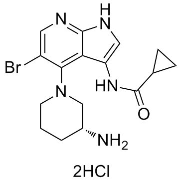
GDC-0575 dihydrochloride is an orally bioavailable CHK1 inhibitor, with an IC50 of 1.2 nM, and has antitumor activity.
|
| Related Catalog |
|
| Target |
Chk1:1.2 nM (IC50)
|
| In Vitro |
GDC-0575 dihydrochloride is a selective and orally bioavailable CHK1 inhibitor, with an IC50 of 1.2 nM. GDC-0575 (100 nM) suppressses CHK1 activation induced by AraC by decreasing the level of Tyr15-phosphorylated CDK2. GDC-0575 (100 nM) has no effect on the viability of AML cells, but significantly reduces cell viability and induces apoptosis in combination with AraC. In addition, GDC-0575 plus AraC shows no effect on normal hematopoietic stem and progenitor cells (HSPCs)[1]. GDC-0575 shows cytotoxic activity against most of 20 melanoma cell lines tested, but several cell lines grown as tumour sphere (TS) are relatively insensitive[2].
|
| In Vivo |
GDC-0575 (7.5 mg/kg, p.o.) in combination with AraC alomost completely eradicates leukemic burden in mice transplanted with U937-Luc cells, and shows more efficient activity than AraC alone. Furthermore, GDC-0575 elevates the cytotoxicity of AraC in different primary AML models in vivo[1]. GDC-0575 (25, 50 mg/kg, p.o.) dose-dependently inhibits the growth of tumor in D20 and C002 xenografts[2].
|
| Cell Assay |
For co-culture experiments, 2 days before initiating the co-culture, feeder cells are plated onto type-I collagen-coated 96-well or 6-well plates and allowed to reach confluence. One day before starting co-culture, they are irradiated at 6.8 Gy and the culture media exchanged. On day 0 of the co-culture, AmL cells are plated at 2 × 10⁵ cells/mL using the correspondent AmL medium. Cells are cultured at 37°C in 5% CO2-humidified incubators at indicated oxygen concentrations. For short-term culture (STC), cells are kept for 1 week in hypoxia (5% O2) with the indicated drugs: 500 nM AraC and/or 100 nM GDC-0575[1].
|
| Animal Admin |
Mice[1] NSG mice are injected intravenously with 1 × 10⁵-106 cells of AmL and 1-3 × 10⁵ cells of hCB CD34+/hBM CD34+. After demonstrating AmL engraftment at 9-11 weeks through FACS analysis of tibia bone marrow aspiration, mice are treated accordingly to proper 7-day treatment regimen with daily 10 mg/kg AraC via subcutaneous injection, 7.5 mg/kg GDC-0575 suspension administered via oral gavage on every other day schedule, and/or 300 μg/kg G-CSF administered daily for 5 days via intraperitoneal injection. One week after the final dosing, mice are killed by cervical dislocation. The femurs, tibias, and pelvis are dissected and flushed with PBS. Red blood cells are lyzed via ammonium chloride. Cells are stained with human-specific FITC-conjugated anti-CD19, PE-conjugated anti-CD33, PE-Cy7-conjugated anti-CD45, and PERCP-conjugated anti-murine CD45 antibodies. Dead cells and debris are excluded via DAPI staining. A BD LSR II flow cytometer is used for analysis. Flow cytometry analysis is performed with FlowJo software. More than 100,000 DAPI-negative events are collected. Engraftment of AmL is said to be present if a single population of mCD45-hCD45+CD33+CD19- cells is present without accompanying mCD45-hCD45+CD33−CD19+cells[1].
|
| References |
[1]. Di Tullio A, et al. The combination of CHK1 inhibitor with G-CSF overrides cytarabine resistance in human acute myeloid leukemia. Nat Commun. 2017 Nov 22;8(1):1679. [2]. Oo ZY, et al. Endogenous Replication Stress Marks Melanomas Sensitive to CHEK1 Inhibitors In Vivo. Clin Cancer Res. 2018 Jun 15;24(12):2901-2912.
|
