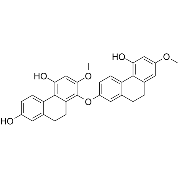
886747-60-0
| Name | Phoyunnanin E |
|---|
| Description | Phoyunnanin E, isolated from Dendrobium venustum, possesses anti-migration activity. Phoyunnanin E can be used for the research of cancer[1]. |
|---|---|
| Related Catalog | |
| In Vitro | Phoyunnanin E (0~10 μM; 48 hours; H460 cells) decreases cell migration through integrin downregulation[1]. Phoyunnanin E (0~100 μM; 24 and 48 hours; H460 cells) shows that the proliferation rate of treated cells have significantly decreased in a dose-dependent manner at the concentration of 10 μM at 48 hours[1]. Phoyunnanin E (0~100 μM; 24 hours; H460 cells) induces cytotoxic effects at the concentrations from 50 to 100 μM and induces a significant increase in apoptotic and necrotic cells at 20~100 μM[1]. Phoyunnanin E (0~10 μM; 48 hours; H460, H292 and A549 cells) significantly decreases the number of cells invading across the matrix and transwell filter within 24 hours in a dose-dependent manner[1]. Phoyunnanin E attenuates anchorage-independent growth and migration of lung cancer H460 cells. Phoyunnanin E significantly inhibits cell migration across the wound space at the concentrations of 5 and 10 μM, at 24 h and 48 h, compared to the non-treated control. Phoyunnanin E (0~10 μM; 48 hours) exhibits a significant decrease in filopodia numbers at the protrusion edges of the cells in a dose-dependent manner. Phoyunnanin E decreases cancer cell migration in other human lung cancer cells. Phoyunnanin E decreases cancer cell invasion. Phoyunnanin E suppresses epithelial-mesenchymal transition (EMT). Phoyunnanin E down-regulates integrins αv and β3 and the regulatory proteins in cell migration[1]. Western Blot Analysis[1] Cell Line: H460 cells Concentration: 1~10 μM Incubation Time: 48 hours Result: Decreased cell migration through integrin downregulation. Cell Proliferation Assay[1] Cell Line: H460 cells Concentration: 0~100 μM Incubation Time: 24 and 48 hours Result: The proliferation rate of treated cells had significantly decreased in a dose-dependent manner at the concentration of 10 μM at 48 h. Apoptosis Analysis[1] Cell Line: H460 cells Concentration: 0~100 μM Incubation Time: 24 hours Result: Induced a significant increase in apoptotic and necrotic cells at 20~100 μM. Cell Cytotoxicity Assay[1] Cell Line: H460 cells Concentration: 0~100 μM Incubation Time: 24 hours Result: Induced cytotoxic effects at the concentrations from 50 to 100 μM. Immunofluorescence[1] Cell Line: H460, H292 and A549 cells Concentration: 0~10 μM Incubation Time: 48 hours Result: Significantly decreased the number of cells invading across the matrix and transwell filter within 24 hours in a dose-dependent manner. |
| References |
| Molecular Formula | C30H26O6 |
|---|---|
| Molecular Weight | 482.524 |
| Storage condition | 2-8℃ |
