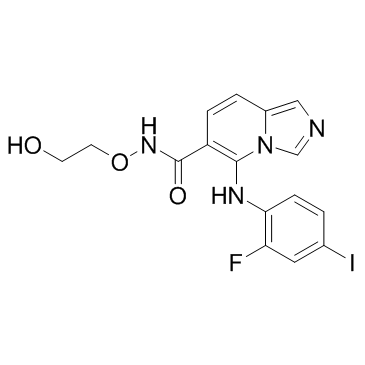
1168091-68-6
| Name | 5-(2-fluoro-4-iodoanilino)-N-(2-hydroxyethoxy)imidazo[1,5-a]pyridine-6-carboxamide |
|---|---|
| Synonyms |
5-[(2-Fluoro-4-iodophenyl)amino]-N-(2-hydroxyethoxy)imidazo[1,5-a]pyridine-6-carboxamide
5-(2-fluoro-4-iodophenylamino)-imidazo[1,5-a]pyridine-6-carboxylic acid (2-hydroxyethoxy)-amide 5-((2-fluoro-4-iodophenyl)amino)-N-(2-hydroxyethoxy)imidazo[1,5-a]pyridine-6-carboxamide GDC-0623 |
| Description | GDC-0623 is a potent, ATP-uncompetitive inhibitor of MEK1 (Ki=0.13 nM, +ATP), and displays 6-fold weaker potency against HCT116 (KRAS (G13D), EC50=42 nM) versus A375 (BRAFV600E, EC50=7 nM). |
|---|---|
| Related Catalog | |
| Target |
MEK1:0.13 nM (Ki, +ATP) |
| In Vitro | DC-0623 and G-573 are able to prevent MEK phosphorylation by CRAF in vitro, and able to block MEK phosphorylation by BRAF(V600E)[1]. GDC-0623 is potent, ATP-uncompetitive inhibitors of MEK1 but shows distinct shifts in cellular activity compared with the other two inhibitors, only 6-fold half-maximum effective concentration (EC50) decreases[2]. |
| In Vivo | GDC-0623 (40 mg/kg, p.o.) shows percent tumour growth inhibition (%TGI) in MiaPaCa-2 xenograft model. GDC-0623 and G-573 show superior antitumour activity compared to GDC-0973 in all three KRAS models[1]. |
| Kinase Assay | 0.14 μM of purified inactive recombinant MEK-1 protein ispreincubated with inhibitors in 15 μL of kinase buffer including (20 mM MOPS pH7.2, 25 mM beta glycerol phosphate, 5 mM EGTA, 1 mM sodium orthovanadate, 1 mM DTT, 100 μM ATP, 15 mM MgCl2). After incubating 10 minutes at 30°C, 1 ng of BRAF, CRAF or BRAF V600E combined with 0.5 μg of inactive recombinant ERK2 isadded to the reaction in total volume of 20 μL. After incubating 30 minutes at 30°C the reaction isstopped by adding LaemmLe sample buffer. Enzyme activity is measured by determining level of phosphor-MEK by SDS-PAGE. Immunoreactive proteins are visualized with SuperSignal West Pico Chemiluminescent Substrate. |
| Cell Assay | Flag-MEK1 mutants, S212P and S212A, are generated using the QuickChange site directed mutagenesis kit. Mammalian expression vectors for N-terminal Flag tagged MEK-1 are expressed in HCT116 cells. 1.8×10⁶ HCT116 cells are plated in 10 cm plate and transfected on the following day with 17 μg of expression constructs using lipofectamine 2000. After 48 hours cells are treated with inhibitors for the indicated times, harvested and lysed in 100 μL cell extraction buffer. Cell lysates from each sample are analyzed by SDS-PAGE. Membranes are incubated with phospho-MEK S221, phospho-ERK1/2 and MEK1 primary antibodies and immunoreactive proteins are analyzed by SuperSignal West Pico Chemiluminescent Substrate. |
| Animal Admin | Colo205 xenografts are established by inoculating 5×10⁶ cells resuspended in Hank's Balanced Salt Solution (HBSS) subcutaneously (s.c.) in the rear right flank of 6-8 week old female nude (nu/nu) mice. NCI-H2122 xenografts are established by inoculating 1×10⁷ cells resuspended in Hank's Balanced Salt Solution (HBSS) plus matrigel (growth factor reduced) s.c. in the rear right flank of 6-8 week old female nu/nu mice. Both A375 and MiaPaca-2 xenografts are initiated by transplanting 1 mm3 tumor fragments from their respective passaged tumors s.c. into the flank of athymic nu/nu mice. When tumors reached appr 200 mm3, mice are randomized and treated with daily (QD) oral gavage (PO) with either vehicle [methylcellulose 0.1% tween 80 0.1% (MCT)], GDC-0973 (at 10 mg/kg), GDC-0623 (40 mg/kg), or G-573 (100 mg/kg). All doses of MEK inhibitors represented maximal tolerated doses (MTDs), resulting in no more than 15-20% body weight loss. Tumor volumes are determined using digital calipers using the formula (L×W×W)/2. Tumor growth inhibition (%TGI) iscalculated as the percentage of the area under the fitted curve (AUC) for the respective dose group per day in relation to the vehicle. Animal weights are recorded twice per week and mice are removed from study if body weights dropped ≥20%. Partial responses (PRs) are defined as any tumor demonstrating a ≥ 50% decrease in tumor volume, whereas complete responses (CRs) are defined as any tumor demonstrating 100% reduction in tumor volume at any point during the study. |
| References |
| Density | 1.8±0.1 g/cm3 |
|---|---|
| Molecular Formula | C16H14FIN4O3 |
| Molecular Weight | 456.210 |
| Exact Mass | 456.009460 |
| PSA | 87.89000 |
| LogP | 4.16 |
| Index of Refraction | 1.703 |
| Storage condition | -20℃ |
