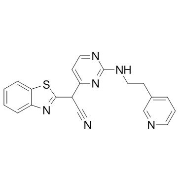
345987-15-7
| Name | 2-(1,3-benzothiazol-2-yl)-2-[2-(2-pyridin-3-ylethylamino)pyrimidin-4-yl]acetonitrile |
|---|---|
| Synonyms |
HMS3265H02
(2s)-1,3-Benzothiazol-2-Yl{2-[(2-Pyridin-3-Ylethyl)amino]pyrimidin-4-Yl}ethanenitrile HMS3265G02 HMS3265G01 HMS3265H01 UNII-Y9A2N9O85G AS601245 |
| Description | AS601245 is a JNK Inhibitor with IC50s of 150, 220, and 70 nM for three JNK human isoforms (hJNK1, hJNK2, and hJNK3), respectively. |
|---|---|
| Related Catalog | |
| Target |
hJNK1:150 nM (IC50) hJNK2:220 nM (IC50) hJNK3:70 nM (IC50) |
| In Vitro | AS601245 inhibits isolated hJNK3 in an ATP-competitive manner. Selectivity of AS601245 is tested against a large panel of kinases. AS601245 exhibits 10- to 20-fold selectivity over c-src, CDK2, and c-Raf and more than 50- to 100-fold selectivity over a range of Ser/Thr- and Tyr-protein kinases[1]. The effects of AS601245 and Clofibrate alone or in association are analysed on proliferation, apoptosis, differentiation and the gene expression profile of CaCo-2 human colon cancer cells. Reduction of cell proliferation, accompanied by the modulation of p21 expression is observed in HepG2 cells, also. 5 μM Clofibrate, 0.1 μM AS601245, and the combined treatment with the two substances do not cause acute toxicity in HepG2 cells. All treatments reduce cell proliferation starting from 48 hours after the beginning of experiments, and the inhibitory effect reaches the maximum at 72 hours[2]. |
| In Vivo | AS601245 is a potent inhibitor of LPS-induced TNF-α release in mice. Administered orally at 0.3, 1, 3, and 10 mg/kg, AS601245 decreases the TNF-α release in a dose-dependent manner. AS601245 (40, 60, and 80 mg/kg) administered i.p. provides significant protection against the delayed loss of hippocampal CA1 neurons in a gerbil model of transient global ischemia. This effect is mediated by JNK inhibition and therefore by c-Jun expression and phosphorylation. A significant neuroprotective effect of AS601245 administered either by i.p. injection (6, 18, and 60 mg/kg) or as i.v. bolus (1 mg/kg) followed by an i.v. infusion (0.6 mg/kg/h) is also observed in rats after focal cerebral ischemia[1]. |
| Kinase Assay | Rat JNK3 assays are performed in 96-well low binding Corning MTT plates: 0.5 μg of recombinant, preactivated GST-JNK3 is incubated with 1 μg of recombinant, biotinylated GST-c-Jun and 2 μM [33Pγ]ATP (2 nCi/μl), in the presence or absence of compounds according to formula I and in a reaction volume of 50 μL containing 50 mM Tris-HCl, pH 8.0; 10 mM MgCl2, 1 mM Dithiothreitol, and 100 μM NaVO4, for 120 min and at room temperature. The reaction is stopped by the addition of 200 μL of a solution containing 250 μg of Streptavidin-coated SPA beads, 5 mM EDTA, 0.1% Triton X-100, and 50 μM ATP, in phosphate saline buffer and further incubation at room temperature for 60 min. After incubation, beads are sedimented by centrifugation at 1500g for 5 min, resuspended in 200 μL of phosphate-buffered saline (PBS) containing 5 mM EDTA, 0.1% Triton X-100, and 50 μM ATP and the radioactivity is measured in a scintillation beta counter, following further sedimentation of the beads by settling down for 60 min at room temperature. Similar method is used to demonstrate inhibition of JNK1 and JNK2[1]. |
| Cell Assay | CaCo-2 cell proliferation is evaluated by using the kit “CellTiter-Glo Luminescent Cell Viability Assay”. This highly sensitive assay detects the luminescence released by the metabolically active cells. Quantification of luminescence is expressed as RLU (relative light unit). For the proliferation experiments, treatments are performed by adding the drugs (e.g., 0.1 μM AS601245) to the CaCo-2 cells seeded at about 4,000 cells/well in a 96-well plate. HepG2 cell proliferation is analysed through the MTT method. Briefly, 1500 cells/well are seeded in 200 μL of serum-supplemented media and the following day treated with the drugs (e.g, 0.1 μM AS601245). 20 μL of 5 mg/mL thiazolyl blue tetrazolium bromide is subsequently added to the cells and removed 2 hours later. 100 μL of DMSO is added to the cells, and the absorbance is recorded at 570 nm through a 96 well plate ELISA reader. Viability is evaluated through Trypan blue exclusion test[2]. |
| Animal Admin | Mice[1] C3H/HEN mice receive an oral treatment with AS601245 (0.3, 1, 3, or 10 mg/kg). Fifteen minutes later, Endotoxins (0.3 mg/kg) are i.p. injected. Heparinized whole blood is collected by retro orbital puncture under isoflurane anesthesia. TNF-α is determined in plasma using an enzyme-linked immunosorbent assay kit. Control animals receive 0.5% CMC/0.25% Tween 20 (10 mL/kg) as vehicle[1]. Rats[1] Wistar rats are randomly divided into a vehicle-treated control group, a reference compound-treated group, and a drug-treated group. The reference compound-treated group receive MK-801 (3 mg/kg i.p.) administered 1 h postischemia onset. The drug-treated group receive AS601245 (6, 18, or 60 mg/kg) administered at the initiation of reperfusion and 5 h later. Control animals receive 0.9% saline (10 ml/kg i.p.). Wistar rats are randomly divided into three groups. The reference compound-treated group receive MK-801 (3 mg/kg i.p.) administered 1 h postischemia onset. The drug-treated group receive an intravenous bolus of AS601245 (1 mg/kg) injected at the initiation of reperfusion followed by an intravenous infusion with a flow of 0.6 mg/kg/h during 22 h. Control animals receive a bolus plus an intravenous infusion of 0.9% saline (10 mL/kg)[1]. |
| References |
| Molecular Formula | C20H16N6S |
|---|---|
| Molecular Weight | 372.44600 |
| Exact Mass | 372.11600 |
| PSA | 118.85000 |
| LogP | 3.21328 |
| Storage condition | 2-8℃ |
