192329-42-3
192329-42-3 structure
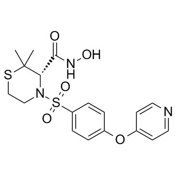
- Name: Prinomastat
- Chemical Name: (3S)-N-hydroxy-2,2-dimethyl-4-(4-pyridin-4-yloxyphenyl)sulfonylthiomorpholine-3-carboxamide
- CAS Number: 192329-42-3
- Molecular Formula: C18H21N3O5S2
- Molecular Weight: 423.50600
- Catalog: Research Areas Cancer
- Create Date: 2018-08-21 18:52:39
- Modify Date: 2025-08-25 11:20:01
-
Prinomastat is a broad spectrum MMP inhibitor with IC50s of 79, 6.3 and 5.0 nM for MMP-1, MMP-3 and MMP-9, respectively.
| Name | (3S)-N-hydroxy-2,2-dimethyl-4-(4-pyridin-4-yloxyphenyl)sulfonylthiomorpholine-3-carboxamide |
|---|---|
| Synonyms |
Prinomastat (USAN/INN)
Prinomastat Prinomastat hydrochloride AG-3354 AG-3362 |
| Description | Prinomastat is a broad spectrum MMP inhibitor with IC50s of 79, 6.3 and 5.0 nM for MMP-1, MMP-3 and MMP-9, respectively. |
|---|---|
| Related Catalog | |
| Target |
IC50: 79 nM (MMP-1), 6.3 nM (MMP-3), 5.0 nM (MMP-9)[1] |
| In Vitro | Co-culture of L/Wnt3a cells and CT7 cells increases the Topflash activity in CT7 cells and this effect is suppressed in the presence of Prinomastat (AG3340). The effect of Wnt1 on the cellular distribution of vimentin is reversed in the presence of Prinomastat in C57MG/Wnt1 cells. Casein gel zymography and western blot analysis of the supernatant of C57MG/Wnt1 cells demonstrate a significant decrease in the presence of MMP-3 in the supernatant of cells treated with Prinomastat or rTIMP-2 that is consistent with these inhibitors inhibiting Wnt1-induced MMP-3 production[3]. |
| In Vivo | Treatment of HaCaT-ras A-5RT3 tumor xenografts with Prinomastat (Ag3340) for 6 days strongly reduces tumor invasion and vascularization. The mean vessel diameter, analyzed by vessel size imaging with MRI, remains constant in untreated animals (day 0: 53±10 μm, day 6: 57±20 μm), but significantly increases in response to Prinomastat treatment (prior to therapy, day 0: 40±10 μm, after therapy, day 6: 70±10 μm; p<0.05). Prinomastat treatment markedly reduces the expression of murine VEGF-A in the stroma of the HaCaT-ras A-5RT3 tumors[2]. |
| Cell Assay | Cells are seeded into 100-mm plates and cultured to confluence in the presence or absence of 10 µg/mL Prinomastat (AG3340). Cells are washed in PBS, resuspended in 70% ethanol and stored overnight at 4°C. Cells are incubated with 20 µg/mL RNase A for 30 min at 37°C, centrifuged, and resuspended in PBS containing 40 µg/mL propidium iodide. The analysis is performed on an cell sorter software[3]. |
| Animal Admin | A total of 14 nude mice are examined using ultrasound (US), starting on day 21 after subcutaneous (s.c.) injection of tumor cells. After the US examination, animals receive either 110 μL MMP inhibitor Prinomastat (Ag3340, 150 mg/kg twice a day, i.p. for 6 days; n=7) or 110 μL NaCl solution (twice a day, i.p. for 6 days; control group, n=7). After 6 days, US examination is repeated. Magnetic resonance imaging (MRI) of treated and untreated animals is only performed at the second time point[2]. |
| References |
| Density | 1.377g/cm3 |
|---|---|
| Molecular Formula | C18H21N3O5S2 |
| Molecular Weight | 423.50600 |
| Exact Mass | 423.09200 |
| PSA | 146.00000 |
| LogP | 4.12300 |
| Index of Refraction | 1.615 |