| Description |
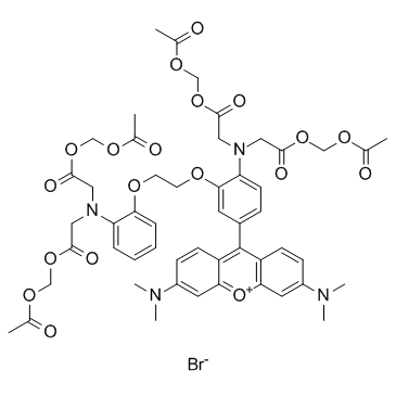
Rhod-2 AM is a fluorescent, mitochondrial probe (λex=552 nm, λem=581 nm).
|
| Related Catalog |
|
| In Vitro |
A high concentration of Ca2+ in mitochondria is illustrated by punctate labeling in cells, which is consistent with the location of Ca2+ in the mitochondria following fluorescence staining with Rhod-2 AM (Rhod2-AM) at 6 h p.i.. Inhibition of mitochondrial Ca2+ uptake by ruthenium red (RR) or 4,4'-diisothiocyanatostilbene-2,2'-disulfonic acid (DIDS) in IMR5 cells infected with poliovirus (PV) is also illustrated following fluorescence staining with Rhod-2 AM at 6 h p.i.[1].
|
| Cell Assay |
For flow cytofluorometry, cells are harvested, pelleted, and resuspended in ice-cold PBS containing 10 mM glucose, 10% fetal bovine serum (FBS), and 10 μM Rhod-2 AM (Rhod2-AM). Mitochondrial calcium levels are determined by the flow cytofluorometry analysis of aliquots of 4×10⁵ cells. For fluorescence microscopy, IMR5 cells are grown on polylysine-coated (10 μg/mL) slides and stained with 7.5 μM Rhod-2 AM in Dulbecco's modified Eagle's medium (DMEM) supplemented with 10% FBS for 2 h before poliovirus (PV) infection. Cells are fixed by incubation for 15 min at 4°C in 4% paraformaldehyde. Cells are washed in PBS, and images are acquired with Zeiss Apotome and Axiovision software[1].
|
| References |
[1]. Brisac C, et al. Calcium flux between the endoplasmic reticulum and mitochondrion contributes to poliovirus-induced apoptosis. J Virol. 2010 Dec;84(23):12226-35.
|


