| Description |
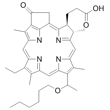
HPPH is a second generation photosensitizer, which acts as a photodynamic therapy (PDT) agent.
|
| Related Catalog |
|
| In Vitro |
Fluorescence image of 4T1 cells incubated with 0.49 µg/mL GO-PEG, 1 μM HPPH (free HPPH) or equivalent amount of GO-PEG-HPPH (1 µM HPPH and 0.49 µg/mL GO-PEG) after 24 h. The cellular uptake of GO-PEG-HPPH and HPPH is investigated with 4T1 murine mammary cancer cells. The cells are incubated with GO-PEG-HPPH and free HPPH at equivalent HPPH concentration (1 µM) for 24 h and then observed with a confocal microscope. Cells treated with GO-PEG-HHPH shows stronger fluorescence signal than those treated with free HPPH. In fact, the fluorescence of HPPH is rather weak[1].
|
| In Vivo |
Tumors are treated with an immune-enhancing PDT regimen followed by a tumor-controlling PDT regimen can leads to enhancement of anti-tumor immunity, while retaining effective control of primary tumor growth. To test this hypothesis, a combination treatment regimen is devised in which Colo26-HA tumor-bearing BALB/c mice are treated with a HPPH-PDT regimen known to lead to enhanced anti-tumor immunity (0.4 μmoles/kg HPPH followed 18 h later by illumination with 665 nm light for a total dose of 48 J/cm2). Following illumination, mice are rested for 9 days; on the ninth day, mice are injected with HPPH. On day 10 following the first treatment, tumors are treated with a tumor control treatment regimen (illumination with 665 nm light for a total dose of 132 J/cm2 given)[2].
|
| Cell Assay |
4T1 cells are cultured in 96-well cell culture plates at 1×10⁴/well for 24 h and then treated with GO-PEG-HPPH, HPPH, or GO-PEG at a series of concentrations (0.078125, 0.15625, 0.3125, 0.625, 1.25, 2.5, 5, 10, and 20 μM). Then, 20 µL of MTT solution (5.0 mg/mL) is added to each well. After the 4 h incubation with the MTT, the media are removed and 100 µL of DMSO is added to solubilize the formazan crystals. The cell toxicity efficacy is measured with a microplate reader at an absorbance of 570 nm[1].
|
| Animal Admin |
Mice[2] Tumor-bearing mice are injected in the tail vein with 0.4 μmol/kg HPPH or 5 mg/kg Porfimer sodium (PII), followed 18-24 h later by illumination to a total light dose of 48 J/cm2 or 132 J/cm2 delivered at a light dose-rate of 14 mW/cm2. Control mice are treated with photosensitizer or light alone. Mice receiving a combination PDT regimen are treated initially with 0.4 μmol/kg HPPH or 5 mg/kg PII followed 18-24 h later by light dose of 48 J/cm2 given at 14 mW/cm2; 9 days later, mice are again injected with photosensitizer and tumors are illuminated with light at a dose of 132 J/cm2 given at 14 mW/cm2[2].
|
| References |
[1]. Rong P, et al. Photosensitizer loaded nano-graphene for multimodality imaging guided tumor photodynamic therapy. Theranostics. 2014 Jan 15;4(3):229-39. [2]. Shams M, et al. Development of photodynamic therapy regimens that control primary tumor growth and inhibit secondary disease. Cancer Immunol Immunother. 2015 Mar;64(3):287-97.
|