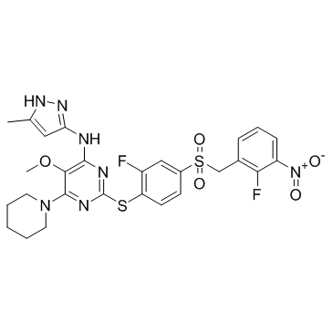
Centrinone-B
Modify Date: 2024-01-08 11:50:02

Centrinone-B structure
|
Common Name | Centrinone-B | ||
|---|---|---|---|---|
| CAS Number | 1798871-31-4 | Molecular Weight | 631.674 | |
| Density | 1.5±0.1 g/cm3 | Boiling Point | 904.9±75.0 °C at 760 mmHg | |
| Molecular Formula | C27H27F2N7O5S2 | Melting Point | N/A | |
| MSDS | N/A | Flash Point | 501.1±37.1 °C | |
Use of Centrinone-BCentrinone-B is a potent and highly selective PLK4 inhibitor, with a Ki of 0.59 nM. |
| Name | Centrinone-B |
|---|---|
| Synonym | More Synonyms |
| Description | Centrinone-B is a potent and highly selective PLK4 inhibitor, with a Ki of 0.59 nM. |
|---|---|
| Related Catalog | |
| Target |
PLK4:0.59 nM (Ki) PLK4 (G95L):497.53 nM (Ki) Aurora A:1239 nM (Ki) Aurora B:5597.14 nM (Ki) |
| In Vitro | Centrinone-B is a potent and highly selective PLK4 inhibitor, with a Ki of 0.59 nM. Centrinone-B slightly binds to Aurora A and Aurora B, with Kis of 1239 nM and 5597.14 nM. Centrinone B exhibits >1000-fold selectivity for Plk4 over Aurora A/B in vitro and does not affect cellular Aurora A or B substrate phosphorylation at concentrations that deplete centrosomes[1]. Centrinone B (0-200 nM) significantly decreases cell viability of PLK4-centriole conjunction melanoma cell lines except p53 mutant SK-MEL-28, and this effect is via inhibition of PLK4. Inhibition of PLK4 by Centrinone B also induces apoptosis in human melanoma cell lines[2]. |
| Kinase Assay | All kinase assays are performed in white 384-well plates. Plk4 assays use equal volumes of: (1) purified 6xHis-tagged human Plk4 kinase domain (aa 2-275) (expressed in E. coli and purified via Ni-NTA affinity chromatography) in 20 mM Tris pH 7.5, 100 mM NaCl, 10% glycerol, 1 mM DTT; (2) 2X reaction buffer consisting of 50 mM HEPES pH 8.5, 20 mM MgCl2, 1 mM DTT, 0.2 mg/mL BSA, 16 μM ATP, and 200 μM A-A11 substrate (amino acid sequence: TPSDSLIYDDGLS). The Plk4 concentration in the final reaction is 2.5-10 nM with a final pH of 8.0. Inhibitors arrayed in dose response are added from DMSO stocks. Reactions are allowed to proceed for 4-16 hours at 25°C. Detection is performed using ADP-Glo reagent. Luminescence is measured on an Infinite M1000 plate reader. Data are fit using Prism and Kis are calculated from IC50 data[1]. |
| Cell Assay | The effect of centrinone B on melanoma cell line and normal melanocyte viability is determined using the CytoTox-Glo assay. Briefly, cells are counted and plated in a 96-well plate and next day, treated with centrinone B for 48 hours, followed by incubation for 15 min with AAF-Glo substrate (alanyl-alanylphenylalanyl-aminoluciferin), which determines a distinct intracellular protease activity related with cytotoxicity (dead-cell protease) via a luminescent signal. Cell viability is determined by subtracting the luminescent signals of dead cells (due to centrinone B) from total dead cells (after addition of digitonin to lyse remaining viable cells). Data are represented as relative light units (RLU) for viable cells[2]. |
| References |
| Density | 1.5±0.1 g/cm3 |
|---|---|
| Boiling Point | 904.9±75.0 °C at 760 mmHg |
| Molecular Formula | C27H27F2N7O5S2 |
| Molecular Weight | 631.674 |
| Flash Point | 501.1±37.1 °C |
| Exact Mass | 631.148315 |
| LogP | 3.77 |
| Vapour Pressure | 0.0±0.3 mmHg at 25°C |
| Index of Refraction | 1.687 |
| Storage condition | 2-8℃ |
| 4-Pyrimidinamine, 2-[[2-fluoro-4-[[(2-fluoro-3-nitrophenyl)methyl]sulfonyl]phenyl]thio]-5-methoxy-N-(5-methyl-1H-pyrazol-3-yl)-6-(1-piperidinyl)- |
| 2-({2-Fluoro-4-[(2-fluoro-3-nitrobenzyl)sulfonyl]phenyl}sulfanyl)-5-methoxy-N-(5-methyl-1H-pyrazol-3-yl)-6-(1-piperidinyl)-4-pyrimidinamine |