BMVC
Modify Date: 2025-10-29 10:11:42
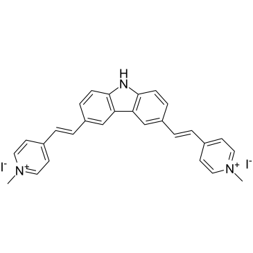
BMVC structure
|
Common Name | BMVC | ||
|---|---|---|---|---|
| CAS Number | 627810-06-4 | Molecular Weight | 657.33 | |
| Density | N/A | Boiling Point | N/A | |
| Molecular Formula | C28H25I2N3 | Melting Point | N/A | |
| MSDS | N/A | Flash Point | N/A | |
Use of BMVCBMVC is a potent G-quadruplex (G4) stabilizer and a selective telomerase inhibitor with an IC50 of ~0.2 μM. BMVC inhibits Taq DNA polymerase with an IC50 of ~2.5 μM. BMVC increases the melting temperature of G4 structure of telomere and accelerates telomere length shortening. Anticancer activities[1][2]. |
| Name | BMVC |
|---|
| Description | BMVC is a potent G-quadruplex (G4) stabilizer and a selective telomerase inhibitor with an IC50 of ~0.2 μM. BMVC inhibits Taq DNA polymerase with an IC50 of ~2.5 μM. BMVC increases the melting temperature of G4 structure of telomere and accelerates telomere length shortening. Anticancer activities[1][2]. |
|---|---|
| Related Catalog | |
| Target |
IC50: ~0.2 μM (Telomerase)[1] G-quadruplex[1] IC50: ~2.5 μM (Taq DNA polymerase)[1] |
| In Vitro | BMVC (0.5 μM; 0-18 days; H1299 cells) treatment markedly increases the percentage of sub-G1-phase cells after 18 days[1]. BMVC (0.5 μM; 0-18 days; H1299 cells) long-term treatment leads to ceasing of cell growth and eventually cell death through apoptosis. The long-term BMVC treatment induces senescence program in H1299 cells[1]. In BMVC-treated cancer cells, hallmarks of senescence, including morphologic changes, detection of senescence-associated β-galactosidase activity, and decreasesd bromodeoxyuridine incorporation, are detected. The BMVC-induced senescence phenotype is accompanied by progressive telomere shortening and detection of the DNA damage foci, indicating that BMVC caused telomere uncapping after long-term treatments[1]. BMVC also suppresses the tumor-related properties of cancer cells, including cell migration, colony-forming ability, and anchorage-independent growth[1]. Cell Cycle Analysis[1] Cell Line: H1299 cells Concentration: 0.5 μM Incubation Time: 0 day, 6 days, 12 days, 18 days Result: The percentage of sub-G1-phase cells was markedly increased after 18 days. Apoptosis Analysis[1] Cell Line: H1299 cells Concentration: 0.5 μM Incubation Time: 0 day, 6 days, 12 days, 18 days Result: Increased apoptotic cells. |
| In Vivo | BMVC (1 mg/kg; intraperitoneal injection; every 3 day; BALB/cAnN.Cg-Foxn1nu/CrlNarl mice) treatment delays tumorigenic potential of cancer cells in vivo[1]. Animal Model: BALB/cAnN.Cg-Foxn1nu/CrlNarl mice injected with H1299 cells[1] Dosage: 1 mg/kg Administration: Intraperitoneal injection; every 3 day Result: The growth rates of tumors in animals were significantly slower than that of control animals. The tumor cells of the mice were indeed entering apoptosis. |
| References |
| Molecular Formula | C28H25I2N3 |
|---|---|
| Molecular Weight | 657.33 |
| InChIKey | FKOQWAUFKGFWLH-UHFFFAOYSA-M |
| SMILES | C[n+]1ccc(C=Cc2ccc3[nH]c4ccc(C=Cc5cc[n+](C)cc5)cc4c3c2)cc1.[I-].[I-] |