| Description |
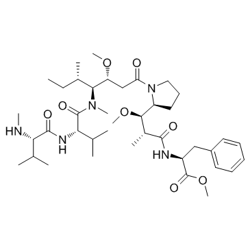
MMAF-Ome belongs to ADC, and inhibits several tumor cell lines with IC50s of 0.056 nM, 0.166 nM, 0.183 nM, and 0.449 nM for MDAMB435/5T4, MDAMB361DYT2, MDAMB468, and Raji (5T4-) cell lines, respectively.
|
| Related Catalog |
|
| In Vitro |
2.5F-Fc and 2.5F-Fc-MMAF have similar IC50 values (6.9±1.1 vs. 8.3±1.3 nM, respectively), indicating that MMAF conjugation has negligible impact on integrin-binding affinity[1].
|
| Cell Assay |
Cells are seeded in a 96-well plate at a density of 2,000 cells per well and grown overnight at 37°C, 5% CO2 in the media described for each cell line above. Cells are subsequently treated with 100 μL of fresh media, containing varying concentrations of knottin-Fc fusion proteins or linker-modified MMAF, and incubated for 5 days at 37°C, 5% CO2. Cell proliferation is measured using the Cell Counting Kit-8 (CCK-8), by adding the water-soluble tetrazolium salt, WST-8, to each well in an amount equal to 10% of the culture volume. After incubation for 1 hour at 37°C, absorbance at 450 nm is measured with a Synergy H4 microtiter plate reader. Cell proliferation is expressed as a percentage of absorbance relative to the control of untreated cells. Percent maximum proliferation is then reported as (sample − background)/(control − background) × 100. Error bars represent the SD of experiments performed in triplicate.
|
| References |
[1]. Currier NV, et al. Targeted Drug Delivery with an Integrin-Binding Knottin-Fc-MMAF Conjugate Produced by Cell-Free Protein Synthesis. Mol Cancer Ther. 2016 Jun;15(6):1291-300.
|
