| 结构式 | 名称/CAS号 | 全部文献 |
|---|---|---|
 |
碘
CAS:7553-56-2 |
|
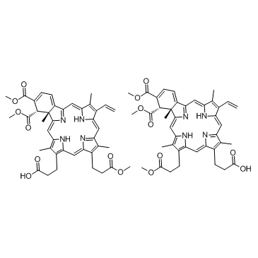 |
维替泊芬
CAS:129497-78-5 |
| 结构式 | 名称/CAS号 | 全部文献 |
|---|---|---|
|
|
碘
CAS:7553-56-2 |
|
|
|
维替泊芬
CAS:129497-78-5 |