| Structure | Name/CAS No. | Articles |
|---|---|---|
 |
Carbon
CAS:7440-44-0 |
|
 |
Hydrochloric acid
CAS:7647-01-0 |
|
 |
Sodium hydroxide
CAS:1310-73-2 |
|
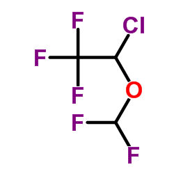 |
Isoflurane
CAS:26675-46-7 |
|
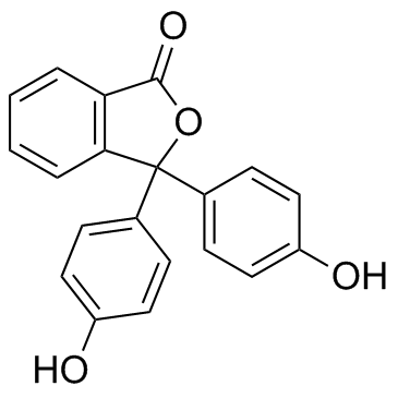 |
Phenolphthalein
CAS:77-09-8 |
|
 |
3-Ethyl-2,4-pentanedione
CAS:1540-34-7 |
|
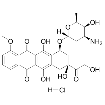 |
Doxorubicin Hydrochloride
CAS:25316-40-9 |
|
 |
HYDROGEN CHLORIDE ~1.25 M IN METHANOL, 250 ML
CAS:132228-87-6 |