MitoTam iodide, hydriodide Suppliers
Total count: 1Updated Date: 2025-02-05 17:55:18

- China(Mainland)
- Contact: Yang
- Phone: 0086-021-52280163
- Email: sales@echemcloud.com
- Website: http://www.echemcloud.com
- Product Name: MitoTam iodide, hydriodide
- Updated Date: 2024-07-15 12:32:41
- Purity: 98.0%
- More information
Inquiry
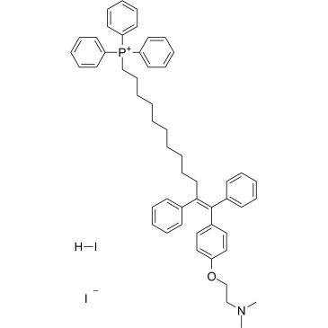
MitoTam iodide, hydriodide
- CAS Number: 1634624-74-0
- Molecular Formula: C52H60I2NOP
- Molecular Weight: 999.82
Related product suppliers
Check more product suppliers