1634624-74-0
1634624-74-0 structure
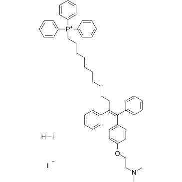
- Name: MitoTam iodide, hydriodide
- Chemical Name: MitoTam iodide, hydriodide
- CAS Number: 1634624-74-0
- Molecular Formula: C52H60I2NOP
- Molecular Weight: 999.82
- Catalog: Signaling Pathways Apoptosis Apoptosis
- Create Date: 2019-09-07 14:53:19
- Modify Date: 2026-06-30 19:09:11
-
MitoTam iodide, hydriodide is a tamoxifen derivative[1], an electron transport chain (ETC) inhibitor, spreduces mitochondrial membrane potential in senescent cells and affects mitochondrial morphology[2].MitoTam iodide, hydriodide is an effective anticancer agent, suppresses respiratory complexes (CI-respiration) and disrupts respiratory supercomplexes (SCs) formation in breast cancer cells[1][2]. MitoTam iodide, hydriodide causes apoptosis[2].
| Name | MitoTam iodide, hydriodide |
|---|
| Description | MitoTam iodide, hydriodide is a tamoxifen derivative[1], an electron transport chain (ETC) inhibitor, spreduces mitochondrial membrane potential in senescent cells and affects mitochondrial morphology[2].MitoTam iodide, hydriodide is an effective anticancer agent, suppresses respiratory complexes (CI-respiration) and disrupts respiratory supercomplexes (SCs) formation in breast cancer cells[1][2]. MitoTam iodide, hydriodide causes apoptosis[2]. |
|---|---|
| Related Catalog | |
| In Vitro | MitoTam (0.5 μM-56 μM; 24 hours) kills breast cancer cell Lines and nonmalignant cells with an IC50 range from 0.65 μM to 55.9 μM[1]. MitoTam (2.5 μM; 2-24 hours) results in stronger activation of the apoptotic pathway in MCF7 Her2high cells compared with mock MCF7 cells[1]. MitoTam (0.05 μM-1 μM; 3 days) causes a concentration-dependent induction of apoptosis in breast cancer cells, while there was no effect for non-malignant breast epithelial cells[2]. Cell Viability Assay[1] Cell Line: Breast Cancer Cell Lines: BT474, MCF7, MCF7 Her2high, MCF7 Her2low, MDA-MB-231, MDA-MB-436, MDA-MB-453, SK-BR-3, T47D; NeuTL cells; Nonmalignant Cells: A014578, H9c2 cells Concentration: 0.5 μM-56 μM Incubation Time: 24 hours Result: Killed breast cancer cells MCF7, MCF7 Her2high, MCF7 Her2low with IC50 values of 1.25 μM, 0.65 μM and 1.45 μM respectively. Western Blot Analysis[2] Cell Line: MCF7 mock and MCF7 Her2high cells Concentration: 2.5 μM Incubation Time: 2 hours, 4 hours, 8 hours, 16 hours, 24 hours Result: Revealed accelerated cleavage of procaspase-9, Parp1/2 and proapoptotic Bax and decreased the antiapoptotic Bcl-2 protein in Her2high cells. Apoptosis Analysis[2] Cell Line: MCF-7, 4T1 and MCF-10a cells Concentration: 0.05 μM-1 μM Incubation Time: 3 days Result: Resulted in apoptosis in MCF7 and 4T1 cells. |
| In Vivo | MitoTam (intraperitoneal injection; 2 μg/g; once a week; 4 weeks) decreases β-gal staining of lungs from MitoTam-treated mice, accompaning by a inhibition in the expression of senescence markers p16Ink4a, p21waf1 and PAI comparing control mice [2]. MitoTam (intraperitoneal injection; 0.54 μmol/mouse; twice a week; 2 weeks) inhibits growth of syngeneic tumors by 80%[1]. MitoTam (intraperitoneal injection; 0.25 μmol/mouse; twice a week; 2 weeks) slows down the growth of MCF7 mock tumors and stops tumor progression after two doses; suppresses Her2high carcinomas decreased threefold from the original size with complete disappearance[1]. Animal Model: 18-month-old or 2-month-old FVB/N mice[1] Dosage: 2 μg/g Administration: Intraperitoneal injection; 2 μg/g; once a week; 4 weeks Result: Eliminated senescent cells also in vivo. Animal Model: 18-month-old or 2-month-old FVB/N mice[2] Dosage: 0.54 μmol/mouse Administration: Intraperitoneal injection; 0.54 μmol/mouse; twice a week; 2 weeks Result: Suppressed Her2high breast carcinomas. Animal Model: 18-month-old or 2-month-old FVB/N mice[1] Dosage: 0.25 μmol/mouse Administration: Intraperitoneal injection; 0.25 μmol/mouse; twice a week; 2 weeks Result: Prevented reaching the ethical endpoint in all situations, slowed down the growth of MCF7 mock tumors and suppressed Her2high carcinomas decreased. |
| References |
| Molecular Formula | C52H60I2NOP |
|---|---|
| Molecular Weight | 999.82 |