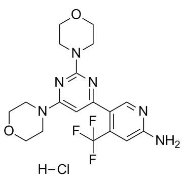
1312445-63-8
| Name | 5-(2,6-dimorpholin-4-ylpyrimidin-4-yl)-4-(trifluoromethyl)pyridin-2-amine hydrochloride |
|---|---|
| Synonyms |
2-Pyridinamine, 5-(2,6-di-4-morpholinyl-4-pyrimidinyl)-4-(trifluoromethyl)-, hydrochloride (1:1)
5-[2,6-Di(4-morpholinyl)-4-pyrimidinyl]-4-(trifluoromethyl)-2-pyridinamine hydrochloride (1:1) NVP-BKM120 Hydrochloride Buparlisib hydrochloride [USAN] BKM 120 hydrochloride 5-(2,6-di-4-morpholinyl-4-pyrimidinyl)-4-trifluoromethylpyridin-2-amine monohydrochloride BUPARLISIB HYDROCHLORIDE BKM120-AAA UNII-194LK4P5K1 5-(2,6-di-4-morpholinylpyrimidinyl)-4-(trifluoromethyl)pyridine-2-amine hydrochloride Buparlisib HCl NVP-BKM120 (Hydrochloride) |
| Description | NVP-BKM120 Hydrochloride is a pan-class I PI3K inhibitor, with IC50 of 52 nM/166 nM/116 nM/262 nM for p110α/p110β/p110δ/p110γ, respectively. |
|---|---|
| Related Catalog | |
| Target |
p110α:52 nM (IC50) p110α-H1047R:58 nM (IC50) p110α-E545K:99 nM (IC50) p110δ:116 nM (IC50) p110β:166 nM (IC50) p110γ:262 nM (IC50) Vps34:2.4 μM (IC50) mTOR:4.6 μM (IC50) |
| In Vitro | NVP-BKM120 (BKM120) exhibits 50-300 nM activity for class I PI3K’s, including the most common p110α mutants. Additionally, NVP-BKM120 exhibits lower potency against class III and class IV PI3K's, where 2, 5, >5, and >25 μM biochemical activity is observed for inhibition of VPS34, mTOR, DNAPK, and PI4K, respectively[1]. NVP-BKM120 induces multiple myeloma (MM) cell apoptosis in both dose- and time-dependent manners. NVP-BKM120 at concentrations ≥10 μM induces significant apoptosis in all tested MM cell lines at 24 h (P<0.05, compares with control). Therefore, 10 μM NVP-BKM120 and 24-h treatment are chose in in the following experiments if not stated otherwise. NVP-BKM120 treatment results in a dose-dependent growth inhibition in all tested MM cell lines. NVP-BKM120 IC50 varies among tested MM cells. At 24 h treatment, IC50 for ARP-1, ARK, and MM.1R is between 1 and 10 μM, while IC50 for MM.1S is <1 μM, and IC50 for U266 is between 10 and 100 μM. In summary, NVP-BKM120 treatment results in MM cell growth inhibition and apoptosis in dose- and time-dependent manners[2]. |
| In Vivo | In A2780 xenograft tumors, oral dosing of NVP-BKM120 (BKM120) at 3, 10, 30, 60, and 100 mg/kg results in a dose dependent modulation of pAKTSer473. Partial inhibition of pAKTSer473 is observed at 3 and 10 mg/kg, and near complete inhibition is observed at doses of 30, 60, or 100 mg/kg, respectively. Inhibition of pAKT (normalized to total AKT) tracked well with both plasma and tumor drug exposure[1]. Mice receiving NVP-BKM120 (5 μM per kg per day for 15 days) treatment has significantly smaller tumor burdens as compare with control mice, which are measured as tumor volume (P<0.05) and level of circulating human kappa chain (P<0.05). In addition, NVP-BKM120 treatment significantly prolongs the survival of tumor-bearing mice (P<0.05)[2]. |
| Kinase Assay | NVP-BKM120 to be tested is dissolved in DMSO and directly distributed into a black 384-well plate at 1.25 μL per well. To start the reaction, 25 μL of 10 nM PI3 kinase and 5 μg/mL 1-alpha-phosphatidylinositol (PI) in assay buffer (10 mM Tris pH 7.5, 5 mM MgCl2, 20 mM NaCl, 1 mM DTT and 0.05% CHAPS) are added into each well followed by 25 μL of 2 μM ATP in assay buffer. The reaction is performed until approx 50% of the ATP is depleted, and then stopped by the addition of 25 μL of KinaseGlo solution. The stopped reaction is incubated for 5 minutes and the remaining ATP is then detected via luminescence[1]. |
| Cell Assay | A2780 cells are cultured in DMEM supplemented with 10% FBS. L-glutamine, sodium pyruvate, and antibiotics. Cells are plated in the same medium at a density of 1000 cells per well, 100 uL per well into black-walled-clear-bottom plates and incubated for 3-5 hours. NVP-BKM120 supplied in DMSO (20 mM) are diluted further into DMSO (7.5 uL of 20 mM NVP-BKM120 in 22.5 uL DMSO. Mix well, transfer 10 uL to 20 uL DMSO, repeat until 9 concentrations have been made). The diluted NVP-BKM120 solution (2 uL), is then added to cell medium (500 uL) cell medium. Equal volumes of this solution (100 uL) are added to the cells in 96 well plates and incubated at 37ºC for 3 days and developed using Cell Titer Glo. Inhibition of cell proliferation is determined by luminescence read using Trilux[1]. |
| Animal Admin | Mice[2] Six- to eight-week-old female severe combined immunodeficiency (SCID) mice are used. SCID mice are subcutaneously inoculated in the right flank with 1 million ARP-1 or MM.1S cells suspended in 50 μL phosphate-buffered saline (PBS). After palpable tumor developed (tumor diameter ≥5 mm), mice are treated with intraperitoneal injection of DMSO/PBS or NVP-BKM120 (5 μM per kg per day) for 15 days. Tumor sizes are measured every 5 days, and blood samples are collected at the same period. Tumor burdens are evaluated by measuring tumor size and detecting circulating human kappa chain or lambda chain. |
| References |
| Molecular Formula | C18H22ClF3N6O2 |
|---|---|
| Molecular Weight | 446.854 |
| Exact Mass | 446.144501 |
| PSA | 90.36000 |
| LogP | 2.67490 |
| Storage condition | 2-8℃ |
| Precursor 4 | |
|---|---|
| DownStream 0 | |


![4,4'-[6-(4,4,5,5-tetramethyl-1,3,2-dioxaborolan-2-yl)pyrimidine-2,4-diyl]di[morpholine] structure](https://image.chemsrc.com/caspic/212/1370351-45-3.png)