5873-16-5
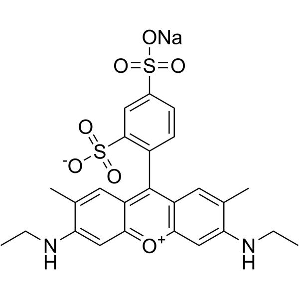
5873-16-5 structure
- Name: hydrogen 9-(2,4-disulphonatophenyl)-3,6-bis(ethylamino)-2,7-dimethylxanthylium, monosodium salt
- Chemical Name: sulforhodamine G
- CAS Number: 5873-16-5
- Molecular Formula: C25H25N2NaO7S2
- Molecular Weight: 552.59500
- Catalog: Dyes and pigments dye Acid Dyestuff
- Create Date: 2018-05-25 08:00:00
- Modify Date: 2026-07-01 21:36:09
-
Sulforhodamine G is a fluorescent stain with broad dynamic ranges. Sulforhodamine G can be used for the research of protein stains[1].
| Name | sulforhodamine G |
|---|---|
| Synonyms |
SULFORHODAMINE G
Xanthylium, 9-(2,4-disulfophenyl)-3,6-bis(ethylamino)-2,7-dimethyl-, inner salt, monosodium salt Sulforhodamine G Dye % sodium 4-[3-(ethylamino)-6-ethylazaniumylidene-2,7-dimethyl-xanthen-9-yl]benzene-1,3-disulfonate 3,6-bis(diethylamino)-2,7-dimethyl-9-(2,4-disulfophenyl)-xanthyl-immonium sodium salt Sulfordamine G EINECS 227-528-6 ACID RED 50 sodium 4-[3-(ethylamino)-6-ethylazaniumylidene-2,7-dimethylxanthen-9-yl]benzene-1,3-disulfonate xanthylium,9-(2,4-disulfophenyl)-3,6-bis(ethylamino)-2,7-dimethyl-,hydroxide Acid Pink G acid red 50 C.I. Acid Red 50 Sulforhodamin G innersalt,monosodiumsalt Sulforhodamine G 9-(2-Sodiosulfo-4-sulfonatophenyl)-3,6-bis(ethylamino)-2,7-dimethylxanthylium C.I.Acid Red 50:C.I sodium 4-[3-(ethylamino)-6-ethyliminio-2,7-dimethyl-xanthen-9-yl]benzene-1,3-disulfonate sodium (Z)-4-(6-(ethylamino)-3-(ethylimino)-2,7-dimethyl-3H-xanthen-9-yl)-3-sulfobenzenesulfonate XANTHYLIUM,9-(2,4-DISULPHOPHENYL)-3,6-BIS(ETHYLAMINO)-2,7 SULFORHODAMINE G ISO 9001:2015 REACH SulphorhodamineG Sulforhodamine G,Acid Red 50 MFCD00213704 |
| Description | Sulforhodamine G is a fluorescent stain with broad dynamic ranges. Sulforhodamine G can be used for the research of protein stains[1]. |
|---|---|
| Related Catalog | |
| In Vitro | Guidelines (Following is our recommended protocol. This protocol only provides a guideline, and should be modified according to your specific needs). Labeling of fluorescent internal protein: A. Prepared of protein samples: 1. Protein samples to be analyzed are spiked with 0.1% of the total protein load of ALIS647 (ALIS internal standard) prior. 2. Protein sample is separated with 2-DE in the dark. B. Purify Sulforhodamine G: 1. Sulforhodamine G (60% purity, 10 mg) dissolved in 100 mL of 1% v/v acetic acid to purify by RP chromatography. 2. Collect the pool with an absorbance maximum at 528 nm, lyophilized to dryness and stored as a dry powder at 47℃. C. Stainning: 1. staining was performed in polypropylene staining dishes wrapped in aluminum foil to prevent photobleaching of the stains. 2. Sulforhodamine G staining is performed overnight in 35% methanol with a four-fold molar excess of dye: protein based on an average protein molecular weight of 50 kDa. 3. Following 4×15 min washes in 35% methanol and 2×15 min equilibrations in water. 4. Total protein and ALIS are visualized with a laser scanner using different channels. 5. Protein spots visualized using total protein stain (λex=532 nm) and ALIS (λex=633 nm) are quantified separately using 2-DE software, and statistical analysis. |
| References |
| Molecular Formula | C25H25N2NaO7S2 |
|---|---|
| Molecular Weight | 552.59500 |
| Exact Mass | 552.10000 |
| PSA | 165.86000 |
| LogP | 6.55920 |
| Personal Protective Equipment | Eyeshields;Gloves;type N95 (US);type P1 (EN143) respirator filter |
|---|---|
| Hazard Codes | F |
| RIDADR | NONH for all modes of transport |
| WGK Germany | 3 |