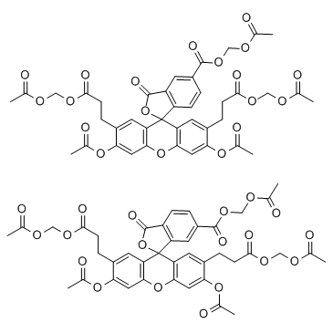
117464-70-7
| Name | 2',7'-Bis(2-carboxyethyl)-5(6)-carboxyfluorescein acetoxymet |
|---|---|
| Synonyms |
Spiro[isobenzofuran-1(3H),9'-[9H]xanthene]-2',7'-dipropanoic acid, 3',6'-bis[(acetyloxy)methoxy]-5-[[(acetyloxy)methoxy]carbonyl]-3-oxo-, bis[(acetyloxy)methyl] ester
MFCD00036969 2',7'-Bis(2-carboxyethyl)-5(6)-carboxyfluorescein tetrakis(acetoxymethyl) ester Acetoxymethyl 3',6'-bis(acetoxymethoxy)-2',7'-bis[3-(acetoxymethoxy)-3-oxopropyl]-3-oxo-3H-spiro[2-benzofuran-1,9'-xanthene]-5-carboxylate 2′,7′-bis(2-Carboxyethyl)-5(6)-carboxyfluorescein acetoxymethyl ester Mixed isomers acetyloxymethyl 3',6'-bis(acetyloxymethoxy)-2',7'-bis[3-(acetyloxymethoxy)-3-oxopropyl]-3-oxospiro[2-benzofuran-1,9'-xanthene]-5-carboxylate BCECF-acetoxymethyl BCECF-AM |
| Description | BCECF-AM is a cell membrane permeable compound, widely used as a fluorescent indicator for intracellular pH. |
|---|---|
| Related Catalog | |
| In Vitro | BCECF-AM is used to measure changes in basal pHi and NHE activity induced by increasing concentrations of ET-1 (0.1-10 nM) in pulmonary arterial smooth muscle cell (PASMC)[1]. |
| Cell Assay | PASMCs are placed in a laminar flow cell chamber perfused with HBSS with pH adjusted to 7.4. pHi is measured in cells incubated with the membrane permeant (acetoxymethyl ester) form of the pH-sensitive fluorescent dye BCECF-AM for 60 min at 37°C under an atmosphere of 20% O2-5% CO2. Cells are then washed with HBSS for 15 min at 37°C to remove extracellular dye and allow complete de-esterification of cytosolic dye. Ratiometric measurement of BCECF fluorescence is performed on a workstation consisting of a Nikon TSE 100 Ellipse inverted microscope with epi-fluorescence attachments. The light beam from a xenon arc lamp is filtered by interference filters at 490 and 440 nm, and focused onto the PASMCS under examination via a 20× fluorescence objective. Light emitted from the cell at 530 nm is returned through the objective and detected by an imaging camera. An electronic shutter is used to minimize photobleaching of dye. Protocols are executed and data collected on-line with InCyte software. pHi is estimated from in situcalibration after each experiment. Cells are perfused with a solution containing (in mM): 105 KCl, 1 MgCl2, 1.5 CaCl2, 10 glucose, 20 HEPES-Tris and 0.01 nigericin to allow pHi to equilibrate to external pH. A two point calibration is created from fluorescence measured as pHi is adjusted with KOH from 6.5 to 7.5. Intracellular H+ ion concentration ([H+]i) is determined from pHi using the formula: pHi = −log ([H+]i). |
| References |
| Density | 1.5±0.1 g/cm3 |
|---|---|
| Boiling Point | 939.2±65.0 °C at 760 mmHg |
| Molecular Formula | C40H36O19 |
| Molecular Weight | 820.70 |
| Flash Point | 368.9±34.3 °C |
| PSA | 264.39000 |
| LogP | 0.90 |
| Vapour Pressure | 0.0±0.3 mmHg at 25°C |
| Index of Refraction | 1.602 |
| Storage condition | -20°C |
| Personal Protective Equipment | dust mask type N95 (US);Eyeshields;Gloves |
|---|---|
| Hazard Codes | Xi: Irritant; |
| Risk Phrases | R36/37/38 |
| Safety Phrases | S22-S26-S36 |
| RIDADR | NONH for all modes of transport |


