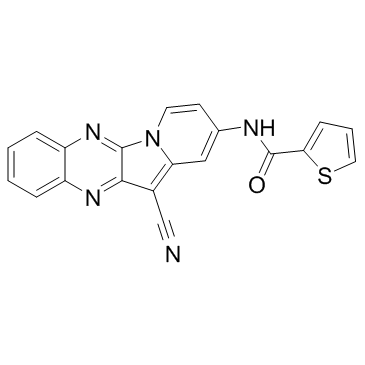
487020-03-1
| Name | N-(12-Cyanindolizino[2,3-b]quinoxalin-2-yl)-2-thiophenecarboxamide |
|---|---|
| Synonyms |
HI TOPK 032
HI-TOPK-032 N-(12-cyanoindolizino[2,3-b]quinoxalin-2-yl)thiophene-2-carboxamide N-(12-Cyanoindolizino[2,3-b]quinoxalin-2-yl)-2-thiophenecarboxami de N-(12-Cyanoindolizino[2,3-b]quinoxalin-2-yl)-2-thiophenecarboxamide |
| Description | HI-TOPK-032 is a potent and specific TOPK inhibitor. |
|---|---|
| Related Catalog | |
| In Vitro | HI-TOPK-032 strongly suppresses TOPK kinase activity but has little effect on extracellular signal-regulated kinase 1 (ERK1), c-jun-NH2-kinase 1, or p38 kinase activities. HI-TOPK-032 occupies the ATP-binding site of TOPK and fits the binding site very well. The compound forms hydrogen bonds with GLY83 and ASP151 and has a hydrophobic interaction with LYS30. However, HI-TOPK-032 at the highest concentration (5 μM) also inhibits MEK1 activity by 40%. HI-TOPK-032 also inhibits anchorage-dependent and -independent colon cancer cell growth by reducing ERK-RSK phosphorylation as well as increasing colon cancer cell apoptosis through regulation of the abundance of p53, cleaved caspase-7, and cleaved PARP[1]. |
| In Vivo | Treatment of mice with 1 or 10 mg/kg of HI-TOPK-032 significantly inhibits HCT-116 tumor growth by more than 60% relative to the vehicle-treated group. Mice are well tolerated with HI-TOPK-032 treatment. The expression of p53 is strongly induced, and phosphorylation of ERK and RSK, a direct downstream protein of ERK, is markedly inhibited in the HI-TOPK-032-treated group[1]. |
| Kinase Assay | The effect of HI-TOPK-032 on ERK1, JNK1, and p38 activity is assessed by an in vitro kinase assay using ERK1 (active, 500 ng), inactive RSK2 (ERK1 substrate, 1 μg), JNK1 (active, 50 ng), c-Jun (JNK1 substrate, 1 μg) and p38 (active, 200 ng), and ATF2 (p38 substrate, 500 ng) with [γ-32P]ATP. Briefly, the reaction is carried out in the presence of 10 μCi of [γ-32P]ATP with HI-TOPK-032 (0.5, 1, 2, 5 μM) in 40 μL of reaction buffer. After incubation at room temperature for 30 minutes, the reaction is stopped by adding 10 μL protein loading buffer and the mixture is separated by SDS-PAGE[1]. |
| Cell Assay | HCT-116 colon cancer cells are treated with different doses of HI-TOPK-032 (1, 2, 5 μM). After incubation for 1, 2, or 3 days, 20 μL of CellTiter96 AQueous One Solution is added and then cells are incubated for 1 hour at 37°C in a 5% CO2 incubator. Absorbance is measured at 492 nm[1]. |
| Animal Admin | Mice: Mice are divided into 4 groups: (i) untreated vehicle group; (ii) 1 mg HI-TOPK-032/kg of body weight; (iii) 10 mg HI-TOPK-032/kg of body weight; and (iv) no cells and 10 mg HI-TOPK-032/kg of body weight. HCT-116 cells are suspended in serum-free McCoy 5A medium and inoculated s.c. into the right flank of each mouse. HI-TOPK-032 or vehicle is injected 3 times per week for 25 days. Tumor volume is calculated[1]. |
| References |
| Density | 1.5±0.1 g/cm3 |
|---|---|
| Boiling Point | 415.3±45.0 °C at 760 mmHg |
| Molecular Formula | C20H11N5OS |
| Molecular Weight | 369.399 |
| Flash Point | 204.9±28.7 °C |
| Exact Mass | 369.068420 |
| PSA | 111.32000 |
| LogP | 2.71 |
| Vapour Pressure | 0.0±1.0 mmHg at 25°C |
| Index of Refraction | 1.808 |
| Storage condition | 2-8℃ |

