552325-16-3
- Name: A-443654
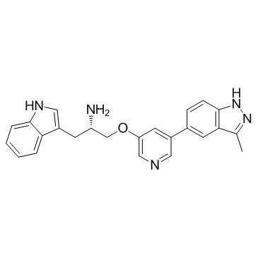
- Chemical Name: (2S)-1-(1H-indol-3-yl)-3-[5-(3-methyl-2H-indazol-5-yl)pyridin-3-yl]oxypropan-2-amine
- CAS Number: 552325-16-3
- Molecular Formula: C24H23N5O
- Molecular Weight: 397.472
- Catalog: Signaling Pathways PI3K/Akt/mTOR Akt
- Create Date: 2016-06-21 11:43:13
- Modify Date: 2026-07-07 18:12:14
-
A-443654 is a potent Akt1/2/3 inhbitor, with a Ki of 160 pM for Akt1.
| Name | (2S)-1-(1H-indol-3-yl)-3-[5-(3-methyl-2H-indazol-5-yl)pyridin-3-yl]oxypropan-2-amine |
|---|---|
| Synonyms |
(2S)-1-(1H-Indol-3-yl)-3-{[5-(3-methyl-1H-indazol-5-yl)-3-pyridinyl]oxy}-2-propanamine
(2s)-1-(1h-Indol-3-Yl)-3-{[5-(3-Methyl-1h-Indazol-5-Yl)pyridin-3-Yl]oxy}propan-2-Amine L20 A-443654 |
| Description | A-443654 is a potent Akt1/2/3 inhbitor, with a Ki of 160 pM for Akt1. |
|---|---|
| Related Catalog | |
| Target |
Akt1:160 pM (Ki) Akt2:160 pM (Ki) Akt3:160 pM (Ki) PKA:6.3 nM (Ki) PKCγ:24 nM (Ki) PKCδ:33 nM (Ki) KDR:3.1 μM (Ki) cKIT:1.2 μM (Ki) SRC:2.6 μM (Ki) Flt1:3.6 μM (Ki) CDK2:24 nM (Ki) ERK2:340 nM (Ki) GSK3β:41 nM (Ki) CK2:2.4 μM (Ki) Chk1:2.3 μM (Ki) RSK2:11 nM (Ki) MAPK-AP2:3.3 μM (Ki) |
| In Vitro | A-443654 exhibits a Ki of 160 pM, a 30,000-fold improvement in potency versus the initial lead molecule. A-443654 is 40-fold selective for Akt over PKA. A-443654 inhibits Akt1, Akt2, or Akt3 equally within cells. A-443654 reduces the P-GSK3 in a dose-responsive manner in all three cell lines. A-443654 inhibits the proliferation of tumor cells with EC50 of 0.1 μM[1]. A-443654-induced morphological changes occur very rapidly (within 2 to 4 h) in both 10A and 10CA1a cells, with 10CA1a cells more sensitive to A-443654 than the 10A cells. A-443654 alone at 2 μM causes the 10CA1a cells, but not the 10A cells, to detach from the plate after 12 h, whereas 1 μM of A-443654 causes 10CA1a cells to detach from the plate after 12 h. FACScan Analysis of rapamycin and A-443654 effects on DNA content in 10A and 10CA1a cells. In contrast, A-443654 at 2 and 5 μM decreases Bcl-2 levels by 30 to 40% in the 10CA1a cells at 8h. The combination of rapamycin with 2 or 5 μM A-443654, however, markedly decreases Bcl-2 protein levels by appr 40 to 50% in the 10A cells and by appr 70% in the 10CA1a cells, respectively[2]. A-443654 demonstrates the greatest selective effect on the mutant cells compared to the WT cells with greater than 3.5 fold relative growth inhibition of the mutant cells[3]. |
| In Vivo | A-443654 (7.5 mg/kg/d, s.c.) inhibits tumor growth in the 3T3-Akt1 flank tumor model. A-443654 (50 mg/kg, s.c.) induces apoptosis in 3T3-Akt1 flank tumors. A-443654 (30 mg/kg, s.c.) leads to increased levels of phosphorylated Akt1 in MiaPaCa-2 tumors[1]. |
| Cell Assay | The cells on 96-well plates are gently washed with 200 μL of PBS. Alamar Blue reagent is diluted 1:10 in normal growth media. The diluted Alamar Blue reagent (100 μL) is added to each well on the 96-well plates and incubated until the reaction is complete as per manufacturer's instructions. Analysis is done using an fmaxFluorescence Microplate Reader, set at the excitation wavelength of 544 nm and emission wavelength of 595 nm. Data are analyzed using SOFTmax PRO software provided by the manufacturer. |
| Animal Admin | Immunocompromised male scid mice are 6 to 8 weeks of age. The 3T3-Akt1 cell line is developed and characterized in our laboratory. The 1×10⁶ 3T3-Akt1 or 2×10⁶ MiaPaCa-2 and PC-3 cells in 50% Matrigel are inoculated s.c. into the flank. For early treatment studies, mice are randomLy assigned to treatment groups and therapy is initiated the day after inoculation. Ten animals are assigned to each group, including controls. For established tumor studies, tumors are allowed to reach a designated size and mice are assigned to treatment groups of equal tumor size (n=10 mice per group). Tumor size is evaluated by twice weekly measurements with digital calipers. Tumor volume is estimated using the formula: V=L×W2/2. A-443654 is given s.c. in a vehicle of 0.2% HPMC. A-674563 is given orally in a vehicle of 5% dextrose. |
| References |
| Density | 1.3±0.1 g/cm3 |
|---|---|
| Boiling Point | 722.0±60.0 °C at 760 mmHg |
| Molecular Formula | C24H23N5O |
| Molecular Weight | 397.472 |
| Flash Point | 390.4±32.9 °C |
| Exact Mass | 397.190247 |
| PSA | 92.61000 |
| LogP | 3.65 |
| Vapour Pressure | 0.0±2.3 mmHg at 25°C |
| Index of Refraction | 1.722 |
| Storage condition | -20°C |