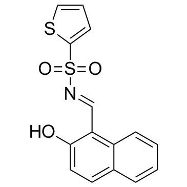
| Description |
STF-083010 is an Ire1 inhibitor. STF-083010 inhibits Ire1 endonuclease activity, without affecting its kinase activity, after endoplasmic reticulum stress.
|
| Related Catalog |
|
| Target |
Ire1[1]
|
| In Vitro |
STF-083010 shows cytostatic and cytotoxic activity in a dose- and time-dependent manner. Treatment with STF-083010 shows significant antimyeloma activity in model human multiple myeloma (MM) xenografts. RPMI 8226 human MM cells grown as tumor xenografts are treated in NSG mice. Intraperitoneal injection of STF-083010 alone (day 1, day 8) significantly inhibits the growth of these tumors[1]. STF-083010 is an IRE1α-specific inhibitor. Four pancreatic cancer cell lines (Panc0403, Panc1005, BxPc3, MiaPaCa2) are treated with different combination of Bortezomib (10 or 50 nM) and STF (10 or 50 μM). The normalized isobologram analysis demonstrates synergistic activity between 10 μM STF and either 10 or 50 nM bortezomib in all four cell lines. Moreover, a higher concentration of STF (50 μM) attains synergy after addition of bortezomib either at a concentration of 10 nM when tested against BxPc3 cells, at a concentration of 50 nM against Panc1005 cells, and at either 10 or 50 nM against Panc0403 cells[2]. STF-083010 (50 μM) suppresses the growth of p53-deficient human cancer cells[3].
|
| In Vivo |
Treatment with STF-083010 reduces the viability of HCT116 p53-/- cells by approximately 20% compared with that of HCT116 p53-/-cells. Administration of STF-083010 to tumors induced by HCT116 p53-/- cells significantly reduces tumor volume and weight by 75% and 73% at the endpoint, respectively[3].
|
| Kinase Assay |
The hIre1α protein, containing both Ire1 cytoplasmic kinase and RNase domains, is expressed and purified from baculovirus. Autophosphorylation activity is determined by the addition of 32P-γATP. Endonuclease activity is determined by the addition of radiolabeled HAC1 508-nt RNA substrate synthesized in vitro using α 32P-UTP. STF083010 is incubated with recombinant hIRE1α protein, radiolabeled HAC1 508 nt RNA, and appropriate buffers. Kinase activity and RNAse cleavage products are quantitated by polyacrylamide gel electrophoresis and 32P-γATP or 32P-UTP autoradiography, respectively[1].
|
| Cell Assay |
Cell viability is determined using the MTT method. After treatment with Tunicamycin (Tm), STF-083010 (50 μM), or both, cells are incubated with MTT solution (1 mg/mL) for 2 h. Isopropanol and HCl are added to the final concentrations of 50% and 20 mM, respectively. The optical density at 570 nm is determined using a spectrophotometer using a reference wavelength of 630 nm[3].
|
| Animal Admin |
Mice[3] Each BALB/c nude mouse (male, 5 weeks of age) is subcutaneously inoculated in the right and left hind footpads with 5×10⁶ HCT116 p53+/+ or HCT116 p53-/- cells. Four days later, DMSO or STF-083010 (40 mg/kg) is intraperitoneally administrated every 3 days. Tumors are measured every 5 days, and their volumes are calculated using the equation mm3=(length (mm))×(width (mm))2/2).
|
| References |
[1]. Papandreou I, et al. Identification of an Ire1alpha endonuclease specific inhibitor with cytotoxic activity against human multiple myeloma. Blood. 2011 Jan 27;117(4):1311-4. [2]. Chien W, et al. Selective inhibition of unfolded protein response induces apoptosis in pancreatic cancer cells. Oncotarget. 2014 Jul 15;5(13):4881-94. [3]. Namba T, et al. Loss of p53 enhances the function of the endoplasmic reticulum through activation of the IRE1α/XBP1 pathway. Oncotarget. 2015 Aug 21;6(24):19990-20001.
|

