186611-52-9
186611-52-9 structure
- Name: IC 261
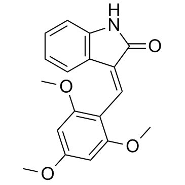
- Chemical Name: (3Z)-3-[(2,4,6-trimethoxyphenyl)methylidene]-1H-indol-2-one
- CAS Number: 186611-52-9
- Molecular Formula: C18H17NO4
- Molecular Weight: 311.332
- Catalog: Signaling Pathways Cell Cycle/DNA Damage Casein Kinase
- Create Date: 2017-10-06 00:18:10
- Modify Date: 2026-07-11 00:38:47
-
IC261 is a selective, ATP-competitive CK1 inhibitor, with IC50s of 1 μM, 1 μM, 16 μM for Ckiδ, Ckiε and Ckiα1, respectively.
| Name | (3Z)-3-[(2,4,6-trimethoxyphenyl)methylidene]-1H-indol-2-one |
|---|---|
| Synonyms |
SU5607
3-[(2,4,6-Trimethoxyphenyl)methylidenyl]-indolin-2-one IC 261 (3E)-3-(2,4,6-Trimethoxybenzylidene)-1,3-dihydro-2H-indol-2-one HMS2230A20 1,3-Dihydro-3-[(2,4,6-trimethoxyphenyl)methylene]-2H-indol-2-one 1,3-Dihydro-3-[(2,4,6-trimethoxyphenyl)methylene]-2H-indol-2-one SU5607 IC-261 IC261 |
| Description | IC261 is a selective, ATP-competitive CK1 inhibitor, with IC50s of 1 μM, 1 μM, 16 μM for Ckiδ, Ckiε and Ckiα1, respectively. |
|---|---|
| Related Catalog | |
| Target |
CKIδ:1 μM (IC50) CKIϵ:1 μM (IC50) CkIα1:16 μM (IC50) |
| In Vitro | IC261 is a selective, ATP-competitive CK1 inhibitor, with IC50s of 1 μM, 1 μM, 16 μM for Ckiδ, Ckiε and Ckiα1, respectively. IC261 is less active on PKA, p34cdc2, and p55fyn (IC50s > 100 μM)[1]. IC261 induces mitotic arrest, spindle defects and centrosome amplification in AC1-M88 cells. IC261 (1 μM) increases G2/M cells after 12 h, and causes cell death at 24 h in AC1-M88 cells. IC261 (1 μM) also induces apoptosis in the extravillous trophoblast hybrid cells[2]. IC261 (1.25 μM) suppresses the proliferation of several pancreatic tumour cell lines, including ASPC-1, BxPc3, Capan-1, Colo357, MiaPaCa-2, Panc1, Panc89, PancTu-1 and PancTu-2 cells. IC261 (1.25 μM) specifically enhances CD95-mediated apoptosis of pancreatic tumour cells[3]. |
| In Vivo | IC261 (20.5 mg/kg) inhibits tumor growth of PancTu-2 cells in SCID mice, downregulates several anti-apoptotic proteins, such as CK1δ/∊, KRAS, and IL6 and upregulates p21, ATM, CHEK1 and STAT1 in mice[3]. |
| Kinase Assay | Casein kinase activity is assayed at 37°C. The standard reaction (40 μL) contains 25 mM 2-(N-morpholino)ethanesulfonic acid, pH 6.5, 50 mM NaCl, 15 mM MgCl2, 2 mg/mL casein, 2 mM EGTA, 100 μM [γ-32P]ATP (100-400 cpm/pmol). Initial velocity measurements are carried out in duplicate with ATP as the varied substrate. Kinetic constants and their standard errors are calculated. For assay of inhibitor potency (IC50), [γ -32P]ATP is held constant (10 μM), whereas IC261 concentration is varied (0.1, 0.3, 1, 3, and 10 μM). To assess kinetic mechanism, inhibitors are held constant (IC261, 20 μM; IC3608, 100 μM), whereas [γ -32P]ATP is varied as above. For screening small molecule libraries, CK1 isoforms (Ckiα1, δ, and ε) are assayed that casein is used at 10 mg/mL, [γ -32P]ATP is held constant at 2 μM or 1 mM[1]. |
| Cell Assay | Human extravillous trophoblast cells irreversibly leave the cell cycle and die when isolated from its natural extracellular matrix. The cell line AC1-M88 is employed in vitro experiments. This cell line is generated by fusion of extravillous trophoblasts with AC1-1. Cells are grown in DMEM (CV-1) or DMEM/F-12 (AC1-M88) medium supplemented with 10% fetal calf serum (FCS) at 37°C in a humidified 5% CO2 atmosphere. Where indicated, cells are γ-irradiated with 5 Gy and harvested at the given time points for western blot analysis, treated with 1 μM IC261 or 0.4 μM nocodazole for 12 h and fixed for immunofluorescence analysis, or treated with 1 μM IC261 and fixed for flow cytometrical analysis or lysed for western blot analysis at the indicated time points. IC261 and nocodazole are dissolved in DMSO as stock solutions (25 and 10 mM, respectively), and control cells are treated with 0.004% DMSO. For immunocytochemistry, the cells are grown on coverslips and are treated with methanol (−20°C) for 5 min, followed by acetone (−20°C) for 20-30 s prior to being used for immunocytochemical detection[2]. |
| Animal Admin | Five million PancTu-1 cells resuspended in 100 µL of a solution containing 50% Matrigel and 50% DMEM/RPMI-1640 (1:1) are injected into the dorsolateral site of 6-week-old C.B-17/IcrHsd-scid-bg mice. After 17 days, mice are randomised to the control group (n = 5), the IC261 treatment group (n = 5), the gemcitabine group (n = 5) and to the IC261/gemcitabine group (n = 5). Injection of dimethylsulfoxide (DMSO; control group), IC261 (20.5 mg/kg), gemcitabine (0.6 mg/kg) alone or in combination (20.5 mg/kg IC261/0.6 mg/kg gemcitabine) (treatment groups) is performed daily for 8 days. Mice are sacrificed by asphyxiation with CO2 the day after the last treatment. Tumours are measured before and during treatment. Finally, the tumours are excised, measured, weighed and fixed in formalin or shock frozen. Tumour volume is calculated according to the formula for a rotational ellipsoid (length × height × width × 0.5236)[3]. |
| References |
| Density | 1.2±0.1 g/cm3 |
|---|---|
| Boiling Point | 555.2±50.0 °C at 760 mmHg |
| Melting Point | 214 °C |
| Molecular Formula | C18H17NO4 |
| Molecular Weight | 311.332 |
| Flash Point | 289.6±30.1 °C |
| Exact Mass | 311.115753 |
| PSA | 60.28000 |
| LogP | 3.01 |
| Vapour Pressure | 0.0±1.5 mmHg at 25°C |
| Index of Refraction | 1.619 |
| Personal Protective Equipment | Eyeshields;Gloves;type N95 (US);type P1 (EN143) respirator filter |
|---|---|
| RIDADR | NONH for all modes of transport |