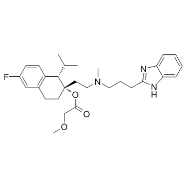
116644-53-2
| Name | [(1S,2S)-2-[2-[3-(1H-benzimidazol-2-yl)propyl-methylamino]ethyl]-6-fluoro-1-propan-2-yl-3,4-dihydro-1H-naphthalen-2-yl] 2-methoxyacetate |
|---|---|
| Synonyms |
Lopac-M-5441
Mibefradil Posicor |
| Description | Mibefradil is a calcium channel blocker with moderate selectivity for T-type Ca2+ channels displaying IC50s of 2.7 μM and 18.6 μM for T-type and L-type currents, respectively. |
|---|---|
| Related Catalog | |
| Target |
IC50: 2.7 μM (T-type calcium channel), 18.6 μM (L-type calcium channel)[1] |
| In Vitro | Mibefradil inhibits reversibly the T- and L-type currents with IC50 values of 2.7 and 18.6 μM, respectively. The inhibition of the L-type current is voltage-dependent, whereas that of the T-type current is not. Ro 40-5967 blocks T-type current already at a holding potential of -100 mV[1] At a higher concentration (20 µM), Mibefradil reduces the amplitude of excitatory junction potentials (by 37±10 %), slows the rate of repolarisation (by 44±16 %) and causes a significant membrane potential depolarisation (from −83±1 mV to −71±5 mV). At a higher Mibefradil concentration (20 µM) there is significant membrane potential depolarisation and a slowing of repolarisation. These actions of Mibefradil are consistent with K+ channel inhibition, which has been shown to occur in human myoblasts and other cells[2]. |
| In Vivo | The hearing thresholds of the 24-26 week old C57BL/6J mice differed following the 4-week treatment period. The hearing threshold at 24 kHz is significantly decreased in the Mibefradil-treated and benidipine-treated groups compared with the saline-treated group (P<0.05)[3]. Compared with the saline-treated group, rats receiving Mibefradil or Ethosuximide show significant lower CaV3.2 expression in the spinal cord and DRG[4]. |
| Animal Admin | Mice[3] A total of 30 male C57BL/6J mice (age, 6-8 weeks) are randomized into three groups for the detection of three calcium channel receptor subunits α1G, α1H and α1I, using reverse transcription-quantitative polymerase chain reaction (RT-qPCR). In addition, a further 30 C57BL/6J male mice (age, 24-26 weeks) are allocated at random into three treatment groups: Saline, Mibefradil and benidipine. Each group is subjected to auditory brainstem recording (ABR) and distortion product otoacoustic emission (DPOAE) tests following treatment. Mibefradil and benidipine are dissolved in physiological saline solution. A preliminary experiment led to the selection of dosages of 30 mg/kg/day Mibefradil and 10 mg/kg/day Benidipine. The drugs are administered to the mice by gavage for four consecutive weeks. Rats[4] Male Sprague-Dawley rats (200-250 g) are used for right L5/6 SNL to induce neuropathic pain. Intrathecal infusion of saline or TCC blockers [Mibefradil (0.7 μg/h) or Ethosuximide (60 μg/h)] is started after surgery for 7 days. Fluorescent immunohistochemistry and Western blotting are used to determine the expression pattern and protein level of CaV3.2. Hematoxylin-eosin and toluidine blue staining are used to evaluate the neurotoxicity of tested agents. |
| References |
| Density | 1.18g/cm3 |
|---|---|
| Boiling Point | 647.6ºC at 760mmHg |
| Molecular Formula | C29H38FN3O3 |
| Molecular Weight | 495.62900 |
| Flash Point | 345.5ºC |
| Exact Mass | 495.29000 |
| PSA | 67.45000 |
| LogP | 5.27090 |
| Vapour Pressure | 1.17E-16mmHg at 25°C |
| Index of Refraction | 1.585 |
| Storage condition | 2-8℃ |
