| Description |
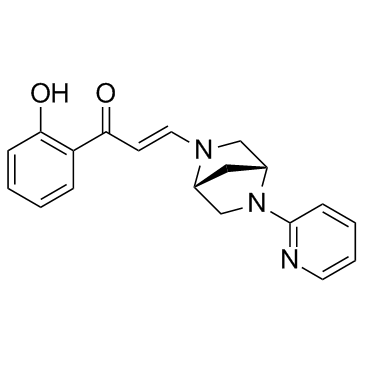
PFI-3 is a selective, potent and cell-permeable SMARCA2/4 bromodomain inhibitor with a Kd of 89 nM.
|
| Related Catalog |
|
| Target |
Kd: 89 nM (SMARCA2/4)[1]
|
| In Vitro |
PFI-3 is a potent, cell-permeable probe capable of displacing ectopically expressed, GFP-tagged SMARCA2-bromodomain from chromatin. PFI-3 binds avidly to both SMARCA2 and SMARCA4 bromodomains (BROMOScan Kd's between 55 and 110 nM) consistent with the binding constant (Kd=89 nM) measured by isothermal titration calorimetry. PFI-3 does not phenocopy the growth inhibitory effects of SMARCA2 knockdown in lung cancer[1]. Exposure of embryonic stem cells to PFI-3 leads to deprivation of stemness and deregulates lineage specification. Furthermore, differentiation of trophoblast stem cells in the presence of PFI-3 is markedly enhanced[2]. PFI-3 binds to certain family VIII bromodomains while displaying significant, broader bromodomain family selectivity. The high specificity of PFI-3 for family VIII is achieved through a novel bromodomain binding mode of a phenolic headgroup that leads to the unusual displacement of water molecules that are generally retained by most other bromodomain inhibitors reported to date[3].
|
| Kinase Assay |
To establish whether PFI-3 intercalates DNA, the compound is assessed using a DNA unwinding assay. PFI-3 (1, 5, or 10 μM), cisplatin, or doxorubicin is incubated with supercoiled pBR322, in the presence of wheat germ topoisomerase I, for 30 min at 37°C. DNA incubated with DMSO in the presence or absence of the enzyme is run as control. After extraction by butanol and chloroform/isoamyl alcohol 24:1, the DNA is run in a 1% (w/v) agarose gel with a 1-kb DNA ladder for 4 hours at 80 V. The gel is then stained with SYBR Safe for 30 min before ultraviolet visualization[2].
|
| References |
[1]. Vangamudi B, et al. The SMARCA2/4 ATPase Domain Surpasses the Bromodomain as a Drug Target in SWI/SNF-Mutant Cancers: Insights from cDNA Rescue and PFI-3 Inhibitor Studies. Cancer Res. 2015 Sep 15;75(18):3865-78. [2]. Fedorov O, et al. Selective targeting of the BRG/PB1 bromodomains impairs embryonic and trophoblast stem cell maintenance. Sci Adv. 2015 Nov 13;1(10):e1500723. [3]. Gerstenberger BS, et al. Identification of a Chemical Probe for Family VIII Bromodomains through Optimization of a Fragment Hit. J Med Chem. 2016 May 26;59(10):4800-11.
|
