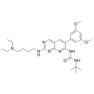
PD173074

PD173074 structure
|
Common Name | PD173074 | ||
|---|---|---|---|---|
| CAS Number | 219580-11-7 | Molecular Weight | 523.670 | |
| Density | 1.2±0.1 g/cm3 | Boiling Point | N/A | |
| Molecular Formula | C28H41N7O3 | Melting Point | 82-85°C | |
| MSDS | Chinese USA | Flash Point | N/A | |
Use of PD173074PD173074 is a potent FGFR1 inhibitor with an IC50 of 25 nM and also inhibits VEGFR2 with an IC50 of 100-200 nM, showing 1000-fold selectivity for FGFR1 over PDGFR and c-Src. |
| Name | 1-tert-butyl-3-[2-[4-(diethylamino)butylamino]-6-(3,5-dimethoxyphenyl)pyrido[2,3-d]pyrimidin-7-yl]urea |
|---|---|
| Synonym | More Synonyms |
| Description | PD173074 is a potent FGFR1 inhibitor with an IC50 of 25 nM and also inhibits VEGFR2 with an IC50 of 100-200 nM, showing 1000-fold selectivity for FGFR1 over PDGFR and c-Src. |
|---|---|
| Related Catalog | |
| Target |
FGFR1:25 nM (IC50) VEGFR2:100 nM (IC50) |
| In Vitro | PD 173074 inhibits autophosphorylation of FGFR1 in a dose-dependent manner with an IC50 in the range 1-5 nM. PD 173074 is an ATP-competitive inhibitor of FGFR1 with an inhibitory constant (Ki) of 40 nM[1]. PD 173074 and SU 5402 produce concentration-dependent reductions in FGF-2 enhancement of granule neuron survival, with IC50 values of 8 nM and 9 μM, respectively. PD 173074 does not inhibit neurotrophic and neuritogenic actions of FGF-2 signalling molecules in cerebellar granule neurons. PD 173074 and SU 5402 concentration-dependently inhibits the neurite growth response, when tested on FGF-2-treated granule neurons growing on polylysine/laminin, with IC50s of 22 nM and 25 μM, respectively[2]. PD173074 effectively antagonizes the effect of FGF-2 on proliferation and differentiation of OL progenitors in culture. Mitogen-activated protein kinase (MAPK) activation, a downstream event after activation of either FGFR or PDGFR, is also blocked by PD173074 in OL progenitors stimulated with FGF-2 but not PDGF[3]. |
| In Vivo | PD 173074 (1 mg/kg, i.p.) exhibits dose-dependent inhibition of FGF-induced neovascularization and angiogenesis in mice[1]. D173074 (25 mg/kg, p.o.) significantly inhibits tumor growth in mice[4]. |
| Cell Assay | An NIH 3T3 cell line overexpressing VEGFR2 (Flk-1) has been described previously. This cell line also expresses FGFR1 endogenously. Cells (1×10⁶) in DMEM supplemented with 10% calf serum are seeded in 10 cm2 dishes and allowed to grow for 48 h. The medium is then removed and the cells are made quiescent in starvation medium (DMEM with 0.1% calf serum). After 18 h, the cells are incubated for 5 min with various concentrations of PD 173074 prepared in starvation medium. The cells are then stimulated with growth factor [VEGF (100 ng/mL) or aFGF (100 ng/mL) and heparin (10 µg/mL)] for 5 min at 37°C. The cells are washed with ice-cold PBS and lysed in 1 mL of lysis buffer (25 mM HEPES pH 7.5, 150 mM NaCl, 1% Triton X-100, 10% glycerol, 1 mM EGTA, 1.5 mM MgCl2, 1 mM PMSF, 10 µg/mL aprotonin, 10 µg/mL leupeptin) containing phosphatase inhibitor (0.2 mM Na3VO4). For inhibition studies of FGFR1, cell lysates are immunoprecipitated with antibodies to FGFR1, and then analyzed by SDS-PAGE and immunoblotting with antibodies to phosphotyrosine. For inhibition studies of VEGFR2, cell lysates (20 µL) are analyzed directly by SDS-PAGE and immunoblotted with antibodies to phosphotyrosine. |
| Animal Admin | Six-week-old athymic nude mice are inoculated subcutaneously with 3×10⁵ NIH 3T3 cells expressing Y373C FGFR3 and Ras V12. Intraperitoneal injections of either 20 mg/kg PD173074 or 0.05 mol/Llactic acid buffer are initiated on the day of tumor injection and continued for 9 days. Ten mice for each experiment are included. |
| References |
| Density | 1.2±0.1 g/cm3 |
|---|---|
| Melting Point | 82-85°C |
| Molecular Formula | C28H41N7O3 |
| Molecular Weight | 523.670 |
| Exact Mass | 523.327087 |
| PSA | 113.53000 |
| LogP | 3.33 |
| Index of Refraction | 1.599 |
| Storage condition | 2-8°C |
| Water Solubility | DMSO: 21 mg/mL |
|
Differentiation of pluripotent stem cells to muscle fiber to model Duchenne muscular dystrophy.
Nat. Biotechnol. , doi:10.1038/nbt.3297, (2015) During embryonic development, skeletal muscles arise from somites, which derive from the presomitic mesoderm (PSM). Using PSM development as a guide, we establish conditions for the differentiation of... |
|
|
Glypican-5 Increases Susceptibility to Nephrotic Damage in Diabetic Kidney.
Am. J. Pathol. 185 , 1889-98, (2015) Type 2 diabetes mellitus is a leading health issue worldwide. Among cases of diabetes mellitus nephropathy (DN), the major complication of type 2 diabetes mellitus, the nephrotic phenotype is often in... |
|
|
Mesenchymal TGF-β signaling orchestrates dental epithelial stem cell homeostasis through Wnt signaling.
Stem Cells 32(11) , 2939-48, (2014) In mouse, continuous growth of the postnatal incisor is coordinated by two populations of multipotent progenitor cells, the dental papilla mesenchymal cells and dental epithelial stem cells, residing ... |
| 1-tert-Butyl-3-[2-{[4-(diethylamino)butyl]amino}-6-(3,5-dimethoxyphenyl)pyrido[2,3-d]pyrimidin-7-yl]urea |
| 1-[2-{[4-(Diethylamino)butyl]amino}-6-(3,5-dimethoxyphenyl)pyrido[2,3-d]pyrimidin-7-yl]-3-(2-methyl-2-propanyl)urea |
| PD173074 |
| PD 173074 |
| Urea, N-[2-[[4-(diethylamino)butyl]amino]-6-(3,5-dimethoxyphenyl)pyrido[2,3-d]pyrimidin-7-yl]-N'-(1,1-dimethylethyl)- |
| 2fgi |