| Structure | Name/CAS No. | Articles |
|---|---|---|
 |
NS-1619
CAS:153587-01-0 |
|
 |
Calphostin C
CAS:121263-19-2 |
|
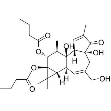 |
Phorbol-12
CAS:37558-16-0 |