NQDI 1
Modify Date: 2025-08-22 15:53:32
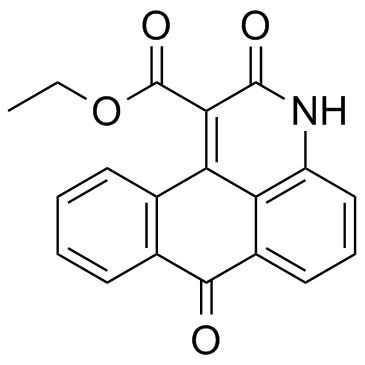
NQDI 1 structure
|
Common Name | NQDI 1 | ||
|---|---|---|---|---|
| CAS Number | 175026-96-7 | Molecular Weight | 319.311 | |
| Density | 1.4±0.1 g/cm3 | Boiling Point | 581.4±50.0 °C at 760 mmHg | |
| Molecular Formula | C19H13NO4 | Melting Point | N/A | |
| MSDS | Chinese USA | Flash Point | 305.4±30.1 °C | |
Use of NQDI 1NQDI-1 inhibits apoptosis signal-regulating kinase 1 (ASK1) with a Ki of 500 nM and an IC50 of 3 μM. |
| Name | Ethyl 2,7-dioxo-2,7-dihydro-3H-naphtho[1,2,3-de]quinoline-1-carbo xylate |
|---|---|
| Synonym | More Synonyms |
| Description | NQDI-1 inhibits apoptosis signal-regulating kinase 1 (ASK1) with a Ki of 500 nM and an IC50 of 3 μM. |
|---|---|
| Related Catalog | |
| Target |
ASK1:3 μM (IC50) |
| In Vitro | The selectivity of NQDI-1 is evaluated in vitro on four serine/threonine protein kinases (protein kinase CK2 (CK2), c-Jun N-terminal kinase 3 (JNK3), Rho-associated protein kinase 1 (Rock1), and Aurora A) and three tyrosine protein kinases (FGFR1, hHGFR, and endothelial TEK tyrosine kinase (Tie2)). The results show that NQDI-1 is a selective inhibitor of ASK1. The activity of FGFR1 protein kinase is inhibited by NQDI-1 (residual activity of 44%). NQDI-1 inhibits ASK1 with a Ki of 500 nM. Inhibition of ASK1 by NQDI-1 is competitive with respect to the phosphodonor substrate ATP[1]. |
| In Vivo | 250 nmol NQDI-1 in DMSO is intracerebroventricularly injected following brain insult. Western blotting is performed to determine the expression of ASK1 in the sham, Hypoxia-ischemia (HI), DMSO and NQDI-1 groups and indicate that NQDI-1 markedly inhibits the expression of ASK1 in the brain cortex, compared with the HI and DMSO group. Furthermore, immunofluorescence staining also indicates that the expression of ASK1 is inhibited by NQDI-1 in the brain cortex. The expression of downstream targets of ASK1 is also determined in the present study. The expression levels of p-JNK, p-c-Jun, p53 and caspase 3 are significantly decreased by NQDI-1, compared with the HI and DMSO groups. Low expression of p-JNK in the brain cortex is also observed by immunofluorescence in the NQDI-1-treated group[2]. |
| Kinase Assay | Enzyme activity of human protein kinases ASK1, Aurora A, ROCK1, HGFR, FGFR1, Tie2, JNK3, and CK2 is determined using in vitro kinase assay (γ-32P-ATP method). Each reaction mixture contains 6 μL of buffer solution (25 mM MOPS, pH 7.2, 2.5 mM EGTA, 2.5 mM EDTA, 0.5 mM DTT, 0.25 mg/mL BSA, 20 mM β-glycerophosphate), 3 μL of substrate solution (MBP, Long S6 kinase substrate peptide, KKKSPGEYVNIEFG, IGF-IRtide (12-527), TK substrate 2, JNK3tide, or RRRDDDSDDD for each kinase, respectively) (5.0 μg/μL), 0.3 μL of enzyme (protein kinase catalytic subunit, 0.1 μg/μL≈32 mU/μL), and 10.25 μL of H2O. The reaction mixture (total volume of 19 μL) is quickly added to 1.5 mL tubes at room temperature. The stock solutions of inhibitors (e.g., NQDI-1) are prepared in DMSO, and the concentration of inhibitor is 1 mM. The concentration of DMSO in the reaction does not exceed 3%. Then 1 μL of inhibitor solution in DMSO is added to each tube and mixed by pipetting. ATP solution is prepared separately. For each sample 0.05 mCi γ-[P32]ATP is taken (specific activity of 100 μCi/μM). The total concentration of labeled and unlabeled ATP is 100 μM. The reaction is started with addition of ATP solution (150 μM ATP, 30 mM MgCl2, 15 mM MOPS, pH 7.2). The time of reaction is 20 min at 30°C. The reaction is stopped by adding 20 μL of 0.5 M orthophosphoric acid. Then the reaction mixture is loaded on the 20 mm filter disks of the cellulose phosphate paper. Filters are washed three times with 0.075 M orthophosphoric acid at room temperature and dried. For detection of products, dried filters are counted by Tri-Carb 2800-TR liquid scintillation analyzer. Then 1 μL of DMSO is added to the reaction volume instead of the inhibitor stock solution for a positive control[1]. |
| Animal Admin | Rats[2] A total of 12 female Sprague-Dawley rats with litters of mixed gender pups are used. The mothers are housed at 25°C under a 12-h light/dark cycle, with ad libitum access to food and water, until the pups are 7-days-old. The HI model is established. The pups are anesthetized with 2.5% halothane and are intracerebroventricularly infused with DMSO or 250 nmol NQDI-1, dissolved in DMSO into the right cerebral hemisphere 30 min prior to HI using a 30-gauge needle with a 5 μL Hamilton syringe (infusion rate, 1 μL/min). |
| References |
| Density | 1.4±0.1 g/cm3 |
|---|---|
| Boiling Point | 581.4±50.0 °C at 760 mmHg |
| Molecular Formula | C19H13NO4 |
| Molecular Weight | 319.311 |
| Flash Point | 305.4±30.1 °C |
| Exact Mass | 319.084473 |
| PSA | 76.49000 |
| LogP | 0.94 |
| Appearance of Characters | light yellow solid |
| Vapour Pressure | 0.0±1.6 mmHg at 25°C |
| Index of Refraction | 1.685 |
| InChIKey | UFJGFNHRMPMALC-UHFFFAOYSA-N |
| SMILES | CCOC(=O)c1c2c3c(cccc3[nH]c1=O)C(=O)c1ccccc1-2 |
| Storage condition | -20℃ |
| RIDADR | NONH for all modes of transport |
|---|

| Precursor 2 | |
|---|---|
| DownStream 0 | |
|
S632A3, a new glutarimide antibiotic, suppresses lipopolysaccharide-induced pro-inflammatory responses via inhibiting the activation of glycogen synthase kinase 3β.
Exp. Cell Res. 318(20) , 2592-603, (2012) Inflammatory mediators including inducible nitric oxide (iNOS), cyclooxygenase-2 (COX-2), tumor necrosis factor-α (TNF-α) and Interleukin-6 (IL-6) contribute to the course of a variety of inflammatory... |
|
|
An ASK1-p38 signalling pathway mediates hydrogen peroxide-induced toxicity in NG108-15 neuronal cells.
Neurosci. Lett. 549 , 163-7, (2013) Reactive oxygen species (ROS) are believed to be involved in many forms of neurodegeneration, including ischaemic infarct damage and Alzheimer's disease. Despite the known involvement of p38 and JNK M... |
|
Name: Primary cell-based high-throughput screening assay for identification of compounds th...
Source: Johns Hopkins Ion Channel Center
Target: regulator of G-protein signaling 4 isoform 2 [Homo sapiens]
External Id: JHICC_RGS_Act_HTS
|
|
Name: Luminescence-based cell-based primary high throughput screening assay to identify ago...
Source: The Scripps Research Institute Molecular Screening Center
Target: mu-type opioid receptor isoform MOR-1 [Homo sapiens]
External Id: OPRM1-OPRD1_AG_LUMI_1536_1X%ACT PRUN
|
|
Name: QFRET-based biochemical primary high throughput screening assay to identify exosite i...
Source: The Scripps Research Institute Molecular Screening Center
Target: disintegrin and metalloproteinase domain-containing protein 17 preproprotein [Homo sapiens]
External Id: ADAM17_INH_QFRET_1536_1X%INH PRUN
|
|
Name: Fluorescence-based cell-based primary high throughput screening assay to identify ago...
Source: The Scripps Research Institute Molecular Screening Center
Target: muscarinic acetylcholine receptor M1 [Homo sapiens]
External Id: CHRM1_AG_FLUO8_1536_1X%ACT PRUN
|
|
Name: uHTS identification of small molecule activators of the adaptive arm of the Unfolded ...
Source: Burnham Center for Chemical Genomics
Target: N/A
External Id: BCCG-A405-UPR-XBP1-PrimaryAgonist-Assay
|
|
Name: High throughput fluorescence intensity-based biochemical assay to screen for small mo...
Source: University of Pittsburgh Molecular Library Screening Center
Target: furin (paired basic amino acid cleaving enzyme), isoform CRA_a [Homo sapiens]
External Id: MH080376 Biochemical HTS for Inhibitors of the Proprotein Convertase Furin.
|
|
Name: Fluorescence polarization to screen for inhibitor that competite the binding of FadD2...
Source: Broad Institute
Target: FATTY-ACID-CoA LIGASE FADD28 (FATTY-ACID-CoA SYNTHETASE)
External Id: 2147-01_Inhibitor_SinglePoint_HTS_Activity
|
|
Name: Bursicon-induced LGR2 mediated cAMP production in LGR-2/CRE6x-Luciferase co-transfect...
Source: Broad Institute
Target: N/A
External Id: Bursicon-induced LGR2 mediated cAMP production in LGR-2/CRE6x-Luciferase co-transfected HEK293 cells Inhibition - 7011-01_Antagonist_SinglePoint_HTS_Activity
|
|
Name: Fluorescence-based cell-based primary high throughput screening assay to identify pos...
Source: The Scripps Research Institute Molecular Screening Center
Target: muscarinic acetylcholine receptor M1 [Homo sapiens]
External Id: CHRM1_PAM_FLUO8_1536_1X%ACT PRUN
|
|
Name: Fluorescence polarization-based biochemical high throughput primary assay to identify...
Source: The Scripps Research Institute Molecular Screening Center
Target: RecName: Full=Sialate O-acetylesterase; AltName: Full=H-Lse; AltName: Full=Sialic acid-specific 9-O-acetylesterase; Flags: Precursor [Homo sapiens]
External Id: SIAE_INH_FP_1536_1X%INH PRUN
|
Total 148, Current Page 1 of 15
1
2
3
4
5
| 3H-Naphtho[1,2,3-de]quinoline-1-carboxylic acid, 2,7-dihydro-2,7-dioxo-, ethyl ester |
| 1-ethyl-anthra<1,9-bc>pyridine-2,7-dione-carboxylate |
| 1-(ethylamino)cyclopentanecarbonitrile |
| Ethyl 2,7-dioxo-2,7-dihydro-3H-naphtho[1,2,3-de]quinoline-1-carboxylate |
| 1-Ethylamino-cyclopentan-carbonsaeure-nitril |
| Cyclopentanecarbonitrile,1-(ethylamino) |
| ethyl 2-hydroxy-7-oxo-7H-naphtho[1,2,3-de]quinoline-1-caboxylate |
| 2-Hydroxy-7-oxo-7H-naphtho[1,2,3-de]chinolin-1-carbonsaeure-aethylester |
| 2-hydroxy-7-oxo-7H-naphtho[1,2,3-de]quinoline-1-carboxylic acid ethyl ester |
| NQDI-1 |