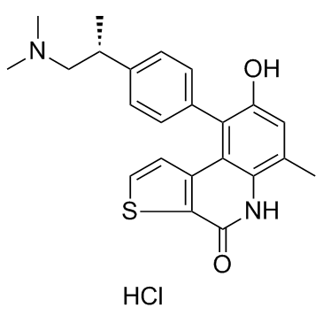
1338545-07-5
| Name | (R)-9-(4-(1-(dimethylamino)propan-2-yl)phenyl)-8-hydroxy-6-methylthieno[2,3-c]quinolin-4(5H)-one hydrochloride |
|---|---|
| Synonyms |
Thieno[2,3-c]quinolin-4(5H)-one, 9-[4-[(1R)-2-(dimethylamino)-1-methylethyl]phenyl]-8-hydroxy-6-methyl-, hydrochloride (1:1)
9-{4-[(2R)-1-(Dimethylamino)-2-propanyl]phenyl}-8-hydroxy-6-methylthieno[2,3-c]quinolin-4(5H)-one hydrochloride (1:1) ots-964 |
| Description | OTS-964 is a potent TOPK inhibitor (IC50=28 nM), which inhibits TOPK kinase activity with high affinity and selectivity. |
|---|---|
| Related Catalog | |
| Target |
IC50: 28 nM (TOPK)[1] |
| In Vitro | OTS-964 (OTS964) inhibits the growth of TOPK-positive cells with low IC50 values [A549 (31 nM), LU-99 (7.6 nM), DU4475 (53 nM), MDA-MB-231 (73 nM), T47D (72 nM), Daudi (25 nM), UM-UC-3 (32 nM), HCT-116 (33 nM), MKN1 (38 nM), MKN45 (39 nM), HepG2 (19 nM), MIAPaca-2 (30 nM), and 22Rv1 (50 nM)], whereas its growth inhibitory effect against TOPK-negative HT29 cancer cells is significantly (P=1.45×10⁻⁴) weaker, with IC50 of 290 nM. Although OTS-964 reveals some suppressive effect on Src family kinases, the response to OTS-964 in these cancer cells is not correlated with the expression of Src family kinases c-Src, Fyn, and Lyn. Treatment with OTS-964 decreases autophosphorylation of TOPK (Thr9), as well as phosphorylation of histone H3 (Ser10), in both T47D and LU-99 cells. Moreover, time lapse imaging in T47D cells shows that treatment with OTS-964 induces cytokinesis defects followed by apoptosis, which is not observed in control DMSO-treated T47D cells[1]. |
| In Vivo | Three LU-99 xenograft mice are intravenously treated with liposomal OTS-964 (40 mg/kg) or vehicle at days 1, 4, 8, and 11, and tumors are collected on day 12. The cellular morphological changes and apoptosis are examined t in the LU-99 cells. Treatment with OTS-964 induced irregular cell morphology with cytokinesis defects. Treatment with OTS-964 significantly increases the number of LU-99 cells with the “intercellular bridge” (P<0.0001), which is one of the markers indicating impaired cell division. The oral administration of free OTS-964 at 50 or 100 mg/kg once every day for 2 weeks results in TGIs of 79 and 113% on day 15, respectively, without any body weight loss. Similar to the intravenous administration of liposomal OTS-964, cell morphological changes, inhibition of TOPK activity, and increased cancer cell death are observed after oral administration of OTS-964 for 1 week. In the 100 mg/kg dosing group, continuous tumor shrinkage is observed after the final administration of the drug, and all six of the mice achieve complete tumor regression (one on day 22, three on day 25, and two on day 29)[1]. |
| Cell Assay | CD34+ HSCs are cultured in RPMI supplemented with 20% fetal bovine serum and 1× StemSpan CC100. Cells are treated with OTS514 (20 or 40 nM) or OTS-964 (100 or 200 nM) for 48 hours. Collected cells are washed with phosphate-buffered saline (PBS) and resuspended in 100 μL of PBS followed by staining with CD41a antibody for 20 min at room temperature. Finally, the cells are washed with PBS again and then analyzed for CD41a staining by flow cytometry on the BD FACSCalibur. Expression of STAT5 is examined by Western blot with an anti-STAT5 antibody[1]. |
| Animal Admin | Mice[1] A549 (1×10⁷ cells) or LU-99 cells (5×10⁶ or 1×10⁷ cells) are injected subcutaneously in the left flank of female BALB/cSLC-nu/nu mice. When A549 xenografts have reached an average volume of 200 mm3 or when LU-99 xenografts have reached an average volume of 150 or 200 mm3, animals are randomized into groups of six mice. The starting tumor volume of 150 mm3 is used for LU-99 xenografts when tumors are monitored for a longer time period (>14 days), because LU-99 cells grow very rapidly, and thus the starting volume of 200 mm3 prevents longer observation considering animal ethics (for example, 200 mm3 of inoculated LU-99 tumor reaches an average tumor volume of about 1100 mm3, whereas A549 tumor reaches about 490 mm3 on day 15). For intravenous administration, three LU-99 xenograft mice are intravenously treated with liposomal OTS-964 (40 mg/kg) into the tail vein or vehicle at days 1, 4, 8, and 11, and tumors are collected on day 12. For oral administration, OTS-964 is treated at 50 or 100 mg/kg once every day for 2 weeks. An administration volume of 10 mL/kg of body weight is used for both administration routes. Concentrations are indicated in the main text and figures. Tumor volumes are determined using a caliper. The weight of the mice is determined as an indicator of tolerability on the same days[1]. |
| References |
| Molecular Formula | C23H25ClN2O2S |
|---|---|
| Molecular Weight | 428.975 |
| Exact Mass | 428.132538 |
| PSA | 84.57000 |
| LogP | 5.89090 |
| Storage condition | -20℃ |


