775294-82-1
775294-82-1 structure
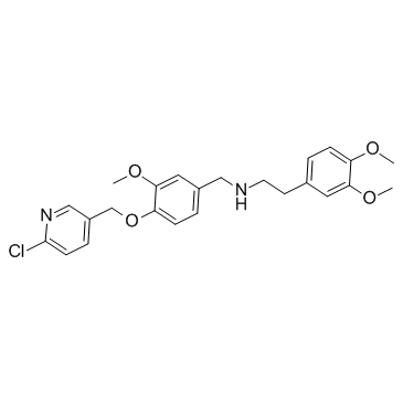
- Name: SBE13
- Chemical Name: SBE13
- CAS Number: 775294-82-1
- Molecular Formula: C24H27ClN2O4
- Molecular Weight: 442.935
- Catalog: Signaling Pathways Autophagy Autophagy
- Create Date: 2018-06-04 23:36:41
- Modify Date: 2026-07-02 12:06:42
-
SBE13 is a potent and selective Plk1 inhibitor, with an IC50 of 200 pM; SBE13 poorly inhibits Plk2 (IC50>66 μM) or Plk3 (IC50=875 nM).
| Name | SBE13 |
|---|---|
| Synonyms |
N-{4-[(6-chloro-3-pyridinyl)methoxy]-3-methoxybenzyl}-N-[2-(3,4-dimethoxyphenyl)ethyl]amine
MFCD05884208 N-{4-[(6-Chloro-3-pyridinyl)methoxy]-3-methoxybenzyl}-2-(3,4-dimethoxyphenyl)ethanamine |
| Description | SBE13 is a potent and selective Plk1 inhibitor, with an IC50 of 200 pM; SBE13 poorly inhibits Plk2 (IC50>66 μM) or Plk3 (IC50=875 nM). |
|---|---|
| Related Catalog | |
| Target |
PLK1:200 pM (IC50) PLK3:875 nM (IC50) |
| In Vitro | SBE13 significantly reduce cell proliferation and induce apoptosis in HeLa cells, with an EC50 of 18 μM[1]. SBE13 (1-100 μM) shows no effect on Caspase 3/7 activity in NIH-3T3 cells. SBE13 (66 and 100 μM) does not change morphology after treatment of primary cells. SBE13 (10 and 100 μM) reduces pRb staining in primary cells, and this indicates a G0/G1 arrest[2]. SBE13 (66 and 100 μM) increases levels of cyclin B1, phospho histone H3, Wee1, Emi1 and securin, and results in cleavage of Cdc27 in HeLa cells. SBE13 (10 and 100 μM) also induces apoptosis of HeLa cells[3]. |
| Kinase Assay | To assay Plk1 kinase activity, cells are lysed after 13 h release in the presence of SBE13 after double thymidine block and kinase is immunoprecipitated from lysates using antibodies. In brief, for each immunoprecipitation 800 μg of total protein are incubated with Plk1 antibody cocktail (1.5 μg) for 2 h at 4°C on a rotator. Immunoprecipitated protein is collected using Protein A/G Agarose beads. Plk1 immunoprecipitates are incubated with casein (1 μg) and with [γ-32P]ATP (1 μCi) for 30 min at 37°C in kinase buffer. Products from the kinase assays are fractionated on 10 % bis-tris-polyacrylamide gels, and phosphorylated substrate is visualized by autoradiography after an exposure of 12-36 h. Equal amounts of immunoprecipitates are subjected to Western blot analysis to confirm equal loading of Plk1 protein in kinase reactions[1]. |
| References |
| Density | 1.2±0.1 g/cm3 |
|---|---|
| Boiling Point | 582.8±45.0 °C at 760 mmHg |
| Molecular Formula | C24H27ClN2O4 |
| Molecular Weight | 442.935 |
| Flash Point | 306.3±28.7 °C |
| Exact Mass | 442.165924 |
| LogP | 3.88 |
| Vapour Pressure | 0.0±1.6 mmHg at 25°C |
| Index of Refraction | 1.577 |
| Storage condition | 2-8℃ |