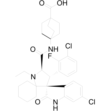
| In Vitro |
APG-115 (0.001-100 μM; 72 hours) inhibits cell proliferation in concentration-dependent manner, with IC50s of 18.9 ± 15.6 nM and 103.5 ± 18.3 nM respectively in AGS and MKN45 cells[3]. APG-115 (0.02 μM, 0.2 μM; 48 hours) enhances the anti-proliferative effect of radiotherapy at different radiation dose[3]. APG-115 (0.02 μM, 0.2 μM; 48 hours) affects progression by inducing cells arrested at G0/G1 phase in AGS and MKN45 cell with wild p53[3]. APG-115 (0.02 μM, 0.2 μM; 24 hours) activates p53 to enhance radiosensitivity in AGS and MKN45 cells; stable knockout of p53 abrogates expression of MDM2, p53, p21, PUMA, BAX, Cleaved-caspase3, γH2AX[3]. APG-115 (0.3 μM, 1 μM, 3 μM, 10 μM; 24 hours) leads to a concentration-dependent cell cycle arrest in G2/M phases and a decreasing in S-phase in p53 wide-type cell lines (TPC-1, KTC-1)[4]. Cell Proliferation Assay[3] Cell Line: AGS and MKN45 cells Concentration: 0.0001 μM, 0.001 μM, 0.01 μM, 0.1 μM, 1 μM, 10 μM, 100 μM Incubation Time: 72 hours Result: Inhibited cell proliferation in a concentration-dependent manner. RT-PCR[3] Cell Line: AGS and MKN45 cells Concentration: 0.02 μM, 0.2 μM Incubation Time: 48 hours Result: Elevated MDM2, p21, PUMA and BAX mRNA expression. Cell Cycle Analysis[3] Cell Line: AGS and MKN45 cells Concentration: 0.02 μM, 0.2 μM Incubation Time: 48 hours Result: Arrested cells at G0/G1 phase. Western Blot Analysis[3] Cell Line: AGS and MKN45 cells Concentration: 0.2 μM Incubation Time: 72 hours Result: Enhanced expressions of MDM2 and p53, stable knockout of p53 abrogated them. Apoptosis Analysis[4] Cell Line: DePTC p53 wide-type cell line: TPC-1 cells, KTC-1 cells Concentration: 0.3μM, 1μM, 3μM, 10 μM Incubation Time: 24 hours Result: Reduced cell population in S-phase, whereas accumulation of cells at G2/M phases.
|
