| In Vitro |
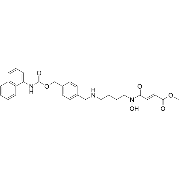
Methylstat (0-5 µM; 48, 72 h) shows anti-proliferative activity with no cytotoxicity on HUVECs at 1-2 µM[1]. Methylstat (0, 1, 2 µM; 48 h) induces cell cycle arrest at G0/G1 phase in a dose-dependent manner[1]. Methylstat (0, 1, 2 µM; 48 h) increases the expression of p53 mRNA levels, the H3K27 methylation levels and the accumulation of p53 and p21 protein levels, but suppresses the protein level of cyclinD1[1]. Methylstat (0, 1, 2 µM) shows anti-angiogenic activity induced by VEGF, bFGF and TNF-α in HUVEC cells, and inhibits the f capillary formation during CAM (chick embryo chorioallantoic membrane) development without any sign of thrombosis and hemorrhage[1]. Methylstat (1.1, 2.2 mM for U266 cells, 2.1, 4.2 mM for ARH77 cells; 72 h) induces apoptosis significantly in U266 and ARH77 cells[2]. Cell Cytotoxicity Assay[1] Cell Line: HUVEC cells Concentration: 0-5 µM Incubation Time: 48, 72 h Result: Did not exhibit cytotoxicity on HUVECs at 1-2 µM. Cell Viability Assay[1] Cell Line: HUVEC, HepG2, HeLa, CHANG cells Concentration: 0-5 µM Incubation Time: 72 h Result: Showed anti-proliferative activity with IC50s of 4, 10, 5, 7.5 µM for HUVEC, HepG2, HeLa, CHANG cells, respectively. Cell Cycle Analysis[1] Cell Line: HUVEC cells Concentration: 0, 1, 2 µM Incubation Time: 48 h Result: G0/G1 phase increased 16.8% compared to non-treated cells, whereas S andG2/M decreased 5.5% and 6.1% respectively. Western Blot Analysis[1] Cell Line: HUVEC cells Concentration: 0, 1, 2 µM Incubation Time: 0-48 h Result: Resulted in accumulation of p53 and p21 protein levels in a time- and dose-dependent manner and increased the H3K27 methylation levels, the but suppressed the protein level of cyclinD1. Apoptosis Analysis[2] Cell Line: U266, ARH77 cells Concentration: 1.1, 2.2 mM for U266 cells, 2.1, 4.2 mM for ARH77 cells Incubation Time: 72 h Result: Induced apoptosis in U266, ARH77 cells.
|
