83280-65-3
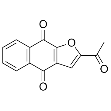
| Name | Napabucasin |
|---|---|
| Synonyms |
2-Acetylnaphtho[2,3-b]furan-4,9-dione
napabucasin BBI608 2-acetylbenzo[f][1]benzofuran-4,9-dione |
| Description | Napabucasin is a STAT3 inhibitor which blocks stem cell activity in cancer cells. |
|---|---|
| Related Catalog | |
| Target |
STAT3 |
| In Vitro | Napabucasin inhibits the expressions of stemness markers and kill stemness-high cancer cells isolated from several kinds of tumors except PCa. Napabucasin not only inhibits cell proliferation, cell motility, cell survival, colony formation ability, and tumorigenic potential of PCa cells, and increases cell apoptosis and sensitivity to docetaxel, but also effectively blocks sphere formation of PrCSCs and kill them as well as inhibits stemness gene expression. Napabucasin inhibits cell proliferation in PC-3 cells and 22RV1 cells at 48, 72, 96, and 120 h (P<0.05). Cell motility and colony formation ability are closely correlated with the process of tumor metastasis. Napabucasin significantly decreases colony formation and cell motility ability of PCa cell lines in vitro (P<0.05). The proliferation of PC-3 and 22RV1 cells treated with 1 μM Napabucasin are significantly decreased from day 2 to 5 compared with the control group (P<0.05)[1]. |
| In Vivo | Napabucasin (40 mg/kg) or Docetaxel significantly reduces xenograft tumor growth and tumor volume (TV) compared with PBS (P<0.05). Notably, while no differences are observed between the Napabucasin and the docetaxel groups in PC-3 mouse xenograft models, the TV in Napabucasin group is even lower than docetaxel group in 22RV1 mouse xenograft models (P<0.05). Additionally, Napabucasin or docetaxel also significantly reduces tumor weight compared with PBS (P<0.05)[1]. |
| Cell Assay | The antiproliferative activity of Napabucasin against the PCa cell lines PC-3 and 22RV1 is examined. For cell proliferation assay, the PCa cell lines (22RV1 and PC-3) are seeded in 96-well plates at 2×10³ cells/well in a final volume of 100 μL and incubated overnight. The proliferation of PC-3 and 22RV1 cells treated with 1 μM Napabucasin. The viability of cells is determined with CellTiter 96 non-radioactive cell proliferation assay (MTS). For colony formation assay, cells are placed in a six-well plate and maintained in RPMI-1640 supplemented with 10% FBS for 2 weeks. The colonies are fixed with 4% paraformaldehyde, stained with 0.1% crystal violet and counted[1]. |
| Animal Admin | Mice[1] A total of 1×10⁶ PC-3 cells or 8×10⁶ 22RV1 cells in 100 μL of PBS are injected subcutaneously into dorsal flanks of an immunodeficient nude mouse. The animals are treated i.p. with Napabucasin (40 mg/kg), Docetaxel (10 mg/kg), or PBS q3d once the tumors have reached 50 mm3. The tumor volume (TV) is calculated every 4 days according to the following standard formula: TV (mm3)=length×width2×0.5. |
| References |
| Density | 1.4±0.1 g/cm3 |
|---|---|
| Boiling Point | 444.4±45.0 °C at 760 mmHg |
| Melting Point | 226 °C |
| Molecular Formula | C14H8O4 |
| Molecular Weight | 240.211 |
| Flash Point | 216.4±21.4 °C |
| Exact Mass | 240.042252 |
| PSA | 64.35000 |
| LogP | 1.68 |
| Vapour Pressure | 0.0±1.1 mmHg at 25°C |
| Index of Refraction | 1.621 |
| Precursor 7 | |
|---|---|
| DownStream 1 | |
![2-(1-hydroxyethyl)benzo[f][1]benzofuran-4,9-dione structure](https://image.chemsrc.com/caspic/268/83889-95-6.png)

