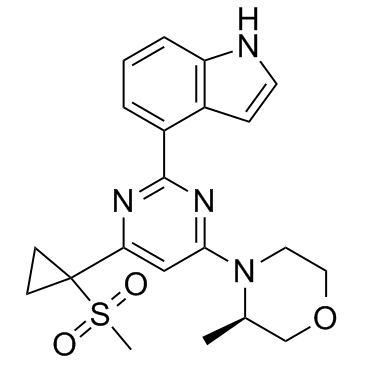
1233339-22-4
| Name | (3R)-4-[2-(3H-indol-4-yl)-6-(1-methylsulfonylcyclopropyl)pyrimidin-4-yl]-3-methylmorpholine |
|---|---|
| Synonyms |
4-{4-[(3R)-3-Methyl-4-morpholinyl]-6-[1-(methylsulfonyl)cyclopropyl]-2-pyrimidinyl}-1H-indole
AZ20 |
| Description | AZ20 is a potent and selective inhibitor of ATR with an IC50 of 5 nM, and has 8-fold selectivity against mTOR (IC50=38 nM). |
|---|---|
| Related Catalog | |
| Target |
ATR:5 nM (IC50) mTOR:38 nM (IC50) PI3Kα:13000 nM (IC50) |
| In Vitro | AZ20 inhibits ATR immunoprecipitated from HeLa nuclear extracts with an IC50 of 5 nM and ATR mediated phosphorylation of Chk1 in HT29 colorectal adenocarcinoma tumor cells with an IC50 of 50 nM[1]. |
| In Vivo | AZ20 (25, 50 mg/kg, p.o.) has high permeability combined with good stability to rat hepatocytes and, despite the lack of progress in achieving markedly higher solubility, has respectable bioavailability in a low dose rat PK study. AZ20 (25, 50 mg/kg, p.o.) leads to significant tumor growth inhibition in female nude mice bearing LoVo tumors[1]. |
| Kinase Assay | ATR for use in the in vitro enzyme assay is obtained from HeLa nuclear extract by immunoprecipitation with rabbit polyclonal antiserum raised to amino acids 400-480 of ATR contained in the following buffer: 25 mM HEPES (pH 7.4), 2 mM MgCl2, 250 mM NaCl, 0.5 mM EDTA, 0.1 mM Na3VO4, 10% v/v glycerol, and 0.01% v/v Tween 20. ATR-antibody complexes are isolated from nuclear extract by incubating with protein A-Sepharose beads for 1 h and then through centrifugation to recover the beads. In the well of a 96-well plate, 10 μL ATR-containing Sepharose beads are incubated with 1 μg of substrate glutathione S-transferase-p53N66 (NH2-terminal 66 amino acids of p53 fused to glutathione S-transferase are expressed in E. coli) in ATR assay buffer (50 mM HEPES (pH 7.4), 150 mM NaCl, 6 mM MgCl2, 4 mM MnCl2, 0.1 mM Na3VO4, 0.1 mM DTT, and 10% (v/v) glycerol) at 37°C in the presence or absence of inhibitor. After 10 min with gentle shaking, ATP is added to a final concentration of 3 μM and the reaction continued at 37°C for an additional 1 h. The reaction is stopped by addition of 100 μL of PBS, and the reaction is transferred to a white opaque glutathione coated 96-well plate and incubated overnight at 4°C. This plate is then washed with PBS/0.05% (v/v) Tween 20, blotted dry, and analyzed by a standard ELISA technique with a phosphoserine 15 p53 antibody. The detection of phosphorylated glutathione S-transferase-p53N66 substrate is performed in combination with a goat anti-mouse horseradish peroxidase-conjugated secondary antibody. Enhanced chemiluminescence solution is used to produce a signal, and chemiluminescent detection is carried out via a TopCount plate reader. The resulting calculated % enzyme activity is then used to determine the IC50 values for the compounds (IC50 taken as the concentration at which 50% of the enzyme activity is inhibited). |
| Cell Assay | Compound dose ranges are created by diluting in 100% DMSO and then further into assay medium (EMEM, 10% FCS, 1% glutamine) using a Labcyte Echo acoustic dispensing instrument. Cells are plated in 384-well Costar plates at 9×10⁴ cells per mL in 40 μL of EMEM, 10% FCS, 1% glutamine and grown for 24 h. Following addition of compound the cells are incubated for 60 min. A final concentration of 3 μM 4NQO (prepared in 100% DMSO) is then added using the Labcyte Echo, and the cells are incubated for a further 60 min. The cells are fixed by adding 40 μL of 3.7% v/v formaldehyde solution for 20 min. After removal of fix, cells are washed with PBS and permeabilized in 40 μL of PBS containing 0.1% Triton X-100. The cells are then washed, and 15 μL primary antibody solution (pChk1 Ser345) is added. The plates are incubated at 4°C overnight. The primary antibody is then washed off, and 20 μL of secondary antibody solution and 1 μM Hoechst 33258 added for 90 min at room temperature. The plates are washed and left in 40 μL of PBS. Plates are then read on an ArrayScan VTI instrument to determine staining intensities, and dose responses are obtained and used to determine the IC50 values for the compounds. |
| Animal Admin | Female Swiss nu/nu mice are housed in negative pressure isolators. LoVo tumor xenografts are established in 8- to 12-week-old mice by injecting 1×10⁷ tumor cells subcutaneously (100 μL in serum free medium) on the left dorsal flank. Animals are randomized into treatment groups when tumors become palpable. AZ20 is prepared in 10% DMSO/40% propylene glycol/50% water and administered orally. Tumors are measured up to three times per week with calipers. Tumor volumes are calculated and the data plotted using the geometric mean for each group versus time. |
| References |
| Density | 1.4±0.1 g/cm3 |
|---|---|
| Boiling Point | 634.6±55.0 °C at 760 mmHg |
| Molecular Formula | C21H24N4O3S |
| Molecular Weight | 412.505 |
| Flash Point | 337.6±31.5 °C |
| Exact Mass | 412.156921 |
| PSA | 93.13000 |
| LogP | 0.50 |
| Vapour Pressure | 0.0±1.9 mmHg at 25°C |
| Index of Refraction | 1.687 |
| Storage condition | -20℃ |
| Symbol |

GHS07 |
|---|---|
| Signal Word | Warning |
| Hazard Statements | H302 |
| Precautionary Statements | P301 + P312 + P330 |
| RIDADR | NONH for all modes of transport |
