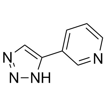
3-TYP
Modify Date: 2024-01-09 14:32:41

3-TYP structure
|
Common Name | 3-TYP | ||
|---|---|---|---|---|
| CAS Number | 120241-79-4 | Molecular Weight | 146.14900 | |
| Density | N/A | Boiling Point | N/A | |
| Molecular Formula | C7H6N4 | Melting Point | N/A | |
| MSDS | N/A | Flash Point | N/A | |
Use of 3-TYP3-TYP is a selective SIRT3 inhibitor, with an IC50 of 16 nM, more potent over SIRT1 (IC50=88 nM), SIRT2 (IC50=92 nM). |
| Name | 3-(1H-1,2,3-triazol-4-yl)pyridine |
|---|---|
| Synonym | More Synonyms |
| Description | 3-TYP is a selective SIRT3 inhibitor, with an IC50 of 16 nM, more potent over SIRT1 (IC50=88 nM), SIRT2 (IC50=92 nM). |
|---|---|
| Related Catalog | |
| Target |
SIRT3:16 nM (IC50) SIRT1:88 nM (IC50) SIRT2:92 nM (IC50) |
| In Vitro | 3-TYP inhibits melatonin-enhanced SIRT3 activity but does not affect SIRT3 protein expression. 3-TYP pretreatment reverses the protective effects of melatonin on cadmium (Cd)-induced mitochondrial-derived O2•− production and autophagic cell death. 3-TYP significantly attenuates melatonin-induced increases in deacetylated-SOD2 expression and SOD2 activity in HepG2 cells exposed to Cd[1]. |
| In Vivo | 3-TYP (50 mg/kg, i.p.) does not significantly influence the LVEF, LVFS, infarct size, serum LDH levels, apoptosis, and oxidative stress compared with those of the Sham group. Moreover, 3-TYP has little effect on gp91phox, Nrf2, NQO 1, Bax, Bcl-2, Caspase-3, and cleaved Caspase-3 expression levels, compared with the Sham group. 3-TYP significantly decreases SIRT3 activity and increases the acetylation of SOD2 compared with that in the control group, without influencing SIRT3 expression. 3-TYP attenuates the cardioprotective effects of melatonin by decreasing the LVEF and LVFS after 24 hour of reperfusion. 3-TYP also increases the infarct size, serum LDH levels, and apoptotic ratio compared with those in the IR+Mel group[2]. |
| Cell Assay | Cell viability is analyzed using Cell Counting Kit-8. Briefly, 1×10⁴ cells are inoculated into 96-well plates. After being treated, 90 μL of medium and 10 μL of CCK-8 solution are added to each well. The cells are then incubated at 37°C for 2 h. After incubation, the absorption at 450 nm is measured using an Infinite™ M200 Microplate Reader. The results are expressed as a percentage of the control. The cell death is also evaluated using the trypan blue assay. HepG2 cells are plated in the 6-well plates (5×10⁵ cells per well) and incubated for 24 h. After being treated with Cd or melatonin, the cells are detached with 300 μL trypsin-EDTA solution. The mixture of detached cells is centrifugated at 300 g for 5 min. Then, the residue is combined with 800 μL trypan blue solution and dispersed. After 3 min staining, cells are counted using an automated cell counter. The dead cells are stained with the blue color. Cell mortality (%) is expressed as percentage of the dead cell number/the total cell number. |
| Animal Admin | In brief, male C57BL/6 mice are anesthetized with 2% isoflurane, and myocardial ischemia is produced by temporarily exteriorizing the heart via a left thoracic incision and placing a 6-0 silk suture slipknot around the left anterior descending coronary artery. After 30 minutes of myocardial ischemia, the slipknot is released, and the myocardium is reperfused for 3 hour (for western blot analysis and oxidative stress measurement) or 24 hour (for cardiac function, apoptotic index and infarct size determination). Sham-operated mice undergo the same surgical procedures except the suture placed under the left coronary artery is not tied. Ten minutes before reperfusion, mice are randomized to receive either vehicle (1% ethanol) or melatonin (20 mg/kg) by intraperitoneal injection. C57BL/6 mice are randomly divided into the following groups: (i) Sham group: mice underwent the sham operation and are treated with vehicle (1% ethanol); (ii) Mel group: mice are treated with melatonin (20 mg/kg via intraperitoneal injection); (iii) IR+V group: mice underwent the MI/R operation and are treated with vehicle (1% ethanol); (iv) IR+Mel group: mice underwent the MI/R operation and are treated with melatonin (20 mg/kg via intraperitoneal injection 10 minutes before reperfusion); (v) IR+Mel+3-TYP group: mice are pretreated with 3-TYP (3-TYP is intraperitoneally injected at a dose of 50 mg/kg every 2 days for a total of three doses prior to the MI/R surgery), subjected to the MI/R operation, and treated with melatonin (20 mg/kg via intraperitoneal injection 10 minutes before reperfusion); and (vi) IR+3-TYP group: mice are pretreated with 3-TYP and then subjected to the MI/R operation. |
| References |
| Molecular Formula | C7H6N4 |
|---|---|
| Molecular Weight | 146.14900 |
| Exact Mass | 146.05900 |
| PSA | 54.46000 |
| LogP | 0.86670 |
| Storage condition | -20℃ |
| 3-(1H-1,2,3-triazol-4-yl) pyridine |
| 3-(1H-[1,2,3]Triazol-4-yl)-pyridine |
| 3-triazolylpyridine |
| 3-(1H-1,2,3-triazol-4-yl)-pyridine |
| 4-(3-Pyridyl)-1,2,3-triazole |
| 3-TYP |