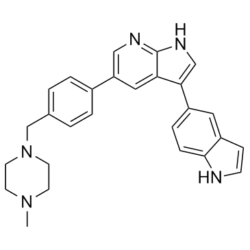
URMC-099

URMC-099 structure
|
Common Name | URMC-099 | ||
|---|---|---|---|---|
| CAS Number | 1229582-33-5 | Molecular Weight | 421.537 | |
| Density | 1.3±0.1 g/cm3 | Boiling Point | N/A | |
| Molecular Formula | C27H27N5 | Melting Point | N/A | |
| MSDS | N/A | Flash Point | N/A | |
Use of URMC-099URMC-099 is an orally bioavailable and potent mixed lineage kinase type 3 (MLK3) (IC50=14 nM) inhibitor with with excellent blood-brain barrier penetration properties. |
| Name | 1H-Pyrrolo[2,3-b]pyridine, 3-(1H-indol-5-yl)-5-[4-[(4-methyl-1-piperazinyl)methyl]phenyl] |
|---|---|
| Synonym | More Synonyms |
| Description | URMC-099 is an orally bioavailable and potent mixed lineage kinase type 3 (MLK3) (IC50=14 nM) inhibitor with with excellent blood-brain barrier penetration properties. |
|---|---|
| Related Catalog | |
| Target |
MLK3:14 nM (IC50) LRRK2:11 nM (IC50) FLT3:4 nM (IC50) FLT1:39 nM (IC50) ABL1 (T315I):3 nM (IC50) ABL1:6.8 nM (IC50) SGK:67 nM (IC50) SGK1:201 nM (IC50) AurA:108 nM (IC50) AurB:123 nM (IC50) AurC:290 nM (IC50) IKKβ:257 nM (IC50) IKKα:591 nM (IC50) TNFα:460 nM (IC50) ROCK1:1030 nM (IC50) ROCK2:111 nM (IC50) CDK1:1125 nM (IC50) CDK2:1180 nM (IC50) TRKA:85 nM (IC50) c-MET:177 nM (IC50) TRKB:217 nM (IC50) IGF1R:307 nM (IC50) LCK:333 nM (IC50) MEKK2:661 nM (IC50) SYK:731 nM (IC50) AMPK:1512 nM (IC50) JNK1:3280 nM (IC50) SRC:4330 nM (IC50) ZAP70:5050 nM (IC50) ERK2:6290 nM (IC50) P38α:12050 nM (IC50) CYP3A4:16.2 μM (IC50) |
| In Vitro | The effect of URMC-099 (URMC099) on the in vitro growth of the “brain homing” MDA-MB-231 BR cells expressing eGFP (eGFP8.4) and their parental cell line, MDA-MB-231 is tested. The cells are treated with either 200 nM URMC-099 or vehicle alone. Cells treated with URMC-099 grow at a similar rate to those treated with vehicle. Cell viability is >99% in all cases[2]. |
| In Vivo | URMC-099 has moderate terminal elimination half-life (t1/2=1.92 h, 2.14 h and 2.72 h for C57 BL/6 mice (10 mg/kg, oral dosing), C57 BL/6 mice (2.5 mg/kg, iv), C57 BL/6 mice (10 mg/kg, iv))[1]. The effect of URMC-099 (URMC099) on tumor formation in vivo is analyzed using a well characterized mouse xenograft model of breast cancer brain metastasis. For these experiments, eGFP8.4 cells are inoculated into the left ventricle of immunodeficient nu/nu mice; animals are then treated with either URMC-099 (10 mg/kg) or vehicle alone, every 12 hours for 20 days. This dose of URMC-099 is chosen because it has been shown to be sufficient to effectively inhibit MLK3 in mice, with good penetration of the blood-brain barrier and potent inhibition of the phosphorylation of Jun N-terminal kinase (JNK) in brain tissue. On day 21 the mice are sacrificed and number of BM is assessed. Fifteen mice are used for each treatment group. BM are detected in 60% of mice, which is consistent with previous studies using this xenograft model by other investigators. URMC-099 treatment significantly (p<0.05, two-tailed t-test) increases the total number of brain metastasis (BM) in mice. For micrometastases, the pattern is similar to that observed for total BM. The number of macrometastases is statistically indistinguishable between mice treated with URMC-099 or vehicle[2]. |
| Cell Assay | MDA-MB-231, MCF10A, HS578t and MDA-MB-231 EGFP8.4 cells are seeded in a 24 well plate at an initial density of 5.0×10⁴ cells/mL in 0.5 mL of media. The cells are treated with either 200 µM of URMC-099 or vehicle (0.002% DMSO). Cell number in each well is measured by trypsinizing the cells and counting them with a hematocytometer. The viability is tested by trypan blue dye exclusion. Each condition is tested in triplicate[2]. |
| Animal Admin | Mice[2] The breast cancer brain metastasis model is performed. Briefly, 6 to 8 week old female nu/nu mice are anesthetized by intraperitoneal injection of 100 mg/kg Ketamine HCl and 10 mg/kg Xylazine, and then inoculated with 100,000 cells in 0.1 mL of cold PBS into the left ventricle. The next day, mice are injected intraperitoneally with URMC-099 at a dose of 10 mg/kg, or vehicle, twice daily for 20 days. On day 21 mice are sacrificed by CO2 suffocation. Brains are removed and fixed with 4% formaldehyde in PBS overnight, then transferred to 30% sucrose in PBS. The brains are then quickly frozen by immersing into isopentane cooled on dry ice. The frozen brains are sectioned coronally every 30 micrometers. Eight sections starting at bregma 2.0 and separated by 360 µm are mounted on glass slides for tumor evaluation under the microscope. The number of brain metastasis (BM) is counted by examining eGFP signals under a fluorescence microscope at 20× magnification[2]. |
| References |
| Density | 1.3±0.1 g/cm3 |
|---|---|
| Molecular Formula | C27H27N5 |
| Molecular Weight | 421.537 |
| Exact Mass | 421.226654 |
| PSA | 50.95000 |
| LogP | 3.85 |
| Index of Refraction | 1.711 |
| Storage condition | 2-8℃ |
| 3-(1H-Indol-5-yl)-5-{4-[(4-methyl-1-piperazinyl)methyl]phenyl}-1H-pyrrolo[2,3-b]pyridine |
| 1H-Pyrrolo[2,3-b]pyridine, 3-(1H-indol-5-yl)-5-[4-[(4-methyl-1-piperazinyl)methyl]phenyl]- |
| URMC-099 |