| Description |
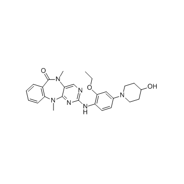
XMD8-92 is a highly selective ERK5/BMK1 inhibitor with dissociation constant (Kd) value of 80 nM.
|
| Related Catalog |
|
| Target |
BMK1:80 nM (Kd)
|
| In Vitro |
XMD8-92 exhibits the greatest affinity towards BMK1 with a measured dissociation constant (Kd) of 80 nM, while DCAMKL2, TNK1 and PLK4 exhibit Kd’s of 190, 890 and 600 nM, respectively. XMD8-92 is profiled against all detectable kinases in HeLa cell lysates using the KiNativ method and is found to be about 10-fold more selective for BMK1 with a IC50 of 1.5 μM than the most potent off-targets, TNK1 (IC50=10 μM) and ACK1 (aka TNK2, IC50=18 μM). Other weak off-targets include the kinase domain 2 of RSK1 and RSK2, PIK4A and PIK4B, and FAK. Notably, MEK5 is not significantly inhibited by XMD8-92 at up to 50 μM[1]. XMD8-92 shows high selectivity to BMK1 in an in vitro ATP-site competition binding assay against 402 kinases as well as in the KiNativ method against all detectable kinases in HeLa cell lysates. XMD8-92 blocks EGF-induced activation of BMK1 with IC50 of 240 nM and, with concentration as high as 5 μM, XMD8-92 has no inhibitory effect on ERK1/2 activation by EGF[2].
|
| In Vivo |
XMD8-92 significantly inhibits tumor growth in vivo by 95%. The pharmacokinetics of XMD8-92 is evaluated in Sprague-Dawley rats given a single intravenous or oral dose. XMD8-92 is found to have a 2.0 hr half life clearance of 26 mL/min/kg. XMD8-92 has moderate tissue distribution with a calculated volume of distribution of 3.4 L/kg. XMD8-92 has high oral bioavailability with 69% of the dose absorbed. After a single oral dose of 2 mg/kg, maximal plasma concentrations of approximately 500 nM are observed by 30 minutes, with 34 nM remaining 8 hr post dose. In tolerability experiments, high plasma concentrations of drug (approximately 10 μM following IP dosing of 50 mg/kg) are maintained throughout the 14 days. XMD8-92 appeares to be well tolerated and the mice appeared healthy with no sign of distress. No vasculature instability is observed in the XMD8-92-treated mice[1]. XMD8-92 treatment in both immunocompetent and immunodeficient mice blocked the growth of lung and cervical xenograft tumors, respectively, by 95%. This remarkable anti-tumor effect of XMD8-92 in lung and cervical xenograft tumor models is due to XMD8-92’s capacity to inhibit tumor cell proliferation through the PML suppression-inducted p21 checkpoint protein, and by blocking of BMK1’s contribution in tumor-associated angiogenesis. Besides, BMK1 knockout (KO) in mice leads to complete and irreversible removal of the BMK1 protein, while XMD8-92 treatment in mice only suppresses the activity of BMK1, which is reversible. Second, the vasculature instability observed in BMK1 KO mice may be due to lack of the C-terminal non-kinase domain of BMK1, which is still present during XMD8-92 treatment of animals[2].
|
| Kinase Assay |
KiNativ profiling of XMD8-92 is carried out with both an ATP and ADP acylphosphate-desthiobiotin with the following modifications. HeLa cell lysates (5 mg/mL total protein) are incubated in the presence of XMD8-92 at 50 μM, 10 μM, 2 μM, 0.8 μM, and 0 μM for 15 minutes prior to addition of the ATP or ADP acylphosphate probe (5 μM final probe concentration). All reactions are performed in duplicate. Probe reactions proceeded for 10 minutes and the reaction stopped by the addition of urea and processed for MS analysis. Samples are analyzed by LC-MS/MS on a linear ion trap mass spectrometer using a time segmented “target list” designed to collect MS/MS spectra from all kinase peptide-probe conjugates that can be detected in HeLa cell lysates. This target list is generated and validated by prior exhaustive analysis of HeLa lysates. Up to four characteristic fragment ions for each kinase peptide-probe conjugate are used to extract signals for each kinase, and a comparison of inhibitor treated to control (untreated) lysates allow for precise determination of % inhibition at each point. A manuscript describing the details of this targeted mass spectrometry approach is in preparation[1].
|
| Animal Admin |
Mice[1] 5×10⁵ HeLa cells are resuspended in DMEM and injected subcutaneously into the right flank of 6-week-old Nod/Scid mice (day 0). On the second day (day 1) after tumor cell injection, mice are randomized into 2 groups (6 animals, XMD8-92, 1-28 day, and 18 animals, control). The XMD8-92 (1-28 day) group is treated with XMD8-92 at the dose of 50 mg/kg twice a day intraperitoneally. The control group receive daily injections of the carrier solution as control. On the day 7, the control group is randomized into 2 groups (6 animals, XMD8-92, 7-28 day, and 12 animals, control). And on the day 14, the remaining control group is randomized into 2 groups (6 animals, XMD8-92, 14-28 day, and 6 animals, control). Treatment with XMD8-92 in XMD8-92 (7-28 day) and XMD8-92 (14-28 day) groups is initiated on day 7 and day 14, respectively. Tumor size is measured using a caliper, and tumor volume is determined[1].
|
| References |
[1]. Yang Q, et al. Pharmacological inhibition of BMK1 suppresses tumor growth through promyelocytic leukemia protein.Cancer Cell. 2010 Sep 14;18(3):258-67. [2]. Yang Q, et al. Targeting the BMK1 MAP kinase pathway in cancer therapy. Clin Cancer Res. 2011 Jun 1;17(11):3527-32. [3]. Umapathy G, et al. The kinase ALK stimulates the kinase ERK5 to promote the expression of the oncogene MYCN in neuroblastoma. Sci Signal. 2014 Oct 28;7(349):ra102.
|
