K-756
Modify Date: 2025-08-20 18:52:57
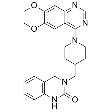
K-756 structure
|
Common Name | K-756 | ||
|---|---|---|---|---|
| CAS Number | 130017-40-2 | Molecular Weight | 433.5 | |
| Density | N/A | Boiling Point | N/A | |
| Molecular Formula | C24H27N5O3 | Melting Point | N/A | |
| MSDS | N/A | Flash Point | N/A | |
Use of K-756K-756 is a direct and selective tankyrase (TNKS) inhibitor, which inhibits the ADP-ribosylation activity of TNKS1 and TNKS2 with IC50s of 31 and 36 nM, respectively. |
| Name | K-756 |
|---|
| Description | K-756 is a direct and selective tankyrase (TNKS) inhibitor, which inhibits the ADP-ribosylation activity of TNKS1 and TNKS2 with IC50s of 31 and 36 nM, respectively. |
|---|---|
| Related Catalog | |
| Target |
TNKS1:31 nM (IC50) TNKS2:36 nM (IC50) |
| In Vitro | K-756 is a novel and selective Wnt/β-catenin pathway inhibitor targeting tankyrase (TNKS). TNKS is one of the members of the PARP family (it is also known as PARP5). K-756 binds to the induced pocket of TNKS and inhibits its enzyme activity. To study the isoform selectivity of K-756, the PARP family enzyme inhibitory activity at 10 μM is evaluated. K-756 inhibits TNKS1 and TNKS2 by 97% and 100%, respectively. In contrast, the inhibitory activity of K-756 against PARP1, PARP2, PARP3, PARP6, PARP7, and PARP11 is less than 13%. K-756 inhibits the cell growth of APC-mutant colorectal cancer COLO 320DM and SW403 cells by inhibiting the Wnt/β-catenin pathway. K-756 strongly inhibits the reporter activity in DLD-1/TCF-Luc cells with an IC50 of 110 nM, but does not inhibit DLD-1/mtTCF-Luc cells, even at 1,000 nM. APC-mutant colorectal cancer cell line COLO 320DM and SW403 cells are treated with K-756 and after 144 hours, cell growth inhibition is measured by an XTT assay. The application of K-756 inhibits the cell growth of COLO 320DM with a GI50 of 780 nM. K-756 also inhibits SW403 with a GI50 of 270 nM[1]. |
| In Vivo | DLD-1/TCF-Luc cell xenografts are created in SCID mice. Vehicle (0.5% MC400) or K-756 is administered orally once a day for 3 days at 100, 200, and 400 mg/kg. The Wnt/β-catenin signal inhibition in the tumor is detected by measuring FGF20 and LGR5 and luciferase activity. The expression of FGF20 and reporter activity are significantly decreased at doses of 100 mg/kg and above at 3-day administration. The expression of LGR5 is significantly decreased at doses of 200 mg/kg and above at 3-day administration. The maximum inhibitory activity is reached with the administration of K-756 at a dose of 400 mg/kg at 3-day administration. The Wnt/β-catenin signal inhibition at a dose of 400 mg/kg is observed from 1-day administration[1]. |
| Kinase Assay | The activity of each enzyme is measured using a PARP1, PARP2, PARP3, PARP6, PARP7, and PARP11 Chemiluminescent Assay Kit, and a TNKS1 and TNKS2 Histone Ribosylation Assay Kit. Enzymatic reactions are conducted in duplicate at 30°C for 1 hour in a 50 μL mixture containing an assay buffer, an enzyme-coated plate, and a substrate. After the enzymatic reactions, 50 μL of Streptavidin-HRP is added to each well and the plate is incubated at room temperature for an additional 30 minutes. 100 μLs of developer reagents are added to the wells and luminescence is measured using a Synergy2 microplate reader. The luminescence data are analyzed using the GraphPad Prism software program[1]. |
| Cell Assay | For the cell viability assay, APC-mutant colorectal cancer cell line COLO 320DM and SW403 cells are seeded in 96-well white plates. siRNAs are reverse transfected by RNAiMax. After 144 hours, a cell viability assay is performed using a Cell Titer-Glo Luminescent Cell Viability Assay Kit. Luciferase activity is measured using a Top count NXT system. For the cell growth inhibition assay, cells are seeded in 96-well plates. The next day, the cells are treated with the compounds (e.g., K-756, 0.1 nM, 1 nM, 10 nM, 100 nM, 1 μM and 10 μM). After 0, 72, or 144 hours of incubation, XTT reagent is added to the cells. After 3 hours of incubation, the formation of formazan dye from tetrazolium salt XTT is measured using a SpectraMax 340PC system[1]. |
| Animal Admin | Mice[1] SCID mice (CB17/Icr-Prkdcscid/CrlCrlj, male, 5 weeks old) are subcutaneously implanted with 5×10⁶ cells of DLD-1/TCF-Luc cells. After 13 days, the mice with a tumor volume of 236.38 to 595.80 mm3 are divided into groups of 5 animals. On the day after grouping, 0.5% MC 400 or K-756 (100, 200, and 400 mg/kg) is orally administered to the mice once a day for 3 days. Twenty-four hours after the last administration of first and second day and 25 hours after the last administration of the third day, the tumor tissues are collected from the mice and frozen in liquid nitrogen. Tumor total RNA is extracted using an RNeasy mini Kit and reverse-transcribed using VILO reagent. An RT-PCR is performed. |
| References |
| Molecular Formula | C24H27N5O3 |
|---|---|
| Molecular Weight | 433.5 |
| InChIKey | GWXCGEJJQFHPPA-UHFFFAOYSA-N |
| SMILES | COc1cc2ncnc(N3CCC(CN4Cc5ccccc5NC4=O)CC3)c2cc1OC |
| Storage condition | 2-8℃ |