5β-Dihydrocortisol
Modify Date: 2025-08-26 17:01:01
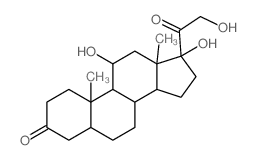
5β-Dihydrocortisol structure
|
Common Name | 5β-Dihydrocortisol | ||
|---|---|---|---|---|
| CAS Number | 1482-50-4 | Molecular Weight | 364.47600 | |
| Density | 1.249g/cm3 | Boiling Point | 544.5ºC at 760mmHg | |
| Molecular Formula | C21H32O5 | Melting Point | N/A | |
| MSDS | N/A | Flash Point | 297.1ºC | |
Use of 5β-Dihydrocortisol5β-Dihydrocortisol, a metabolite of Cortisol, is a potential mineralocorticoid. 5β-Dihydrocortisol can potentiate glucocorticoid activity in raising the intraocular pressure. 5β-Dihydrocortisol causes breast cancer cell apoptosis[1][2][3][4][5]. |
| Name | (8S,9S,10S,13S,14S,17R)-11,17-dihydroxy-17-(2-hydroxyacetyl)-10,13-dimethyl-2,4,5,6,7,8,9,11,12,14,15,16-dodecahydro-1H-cyclopenta[a]phenanthren-3-one |
|---|---|
| Synonym | More Synonyms |
| Description | 5β-Dihydrocortisol, a metabolite of Cortisol, is a potential mineralocorticoid. 5β-Dihydrocortisol can potentiate glucocorticoid activity in raising the intraocular pressure. 5β-Dihydrocortisol causes breast cancer cell apoptosis[1][2][3][4][5]. |
|---|---|
| Related Catalog | |
| In Vitro | 5β-Dihydrocortisol (10-100 μM; 48 h) inhibits the viability of MCF-7 cells, with an IC50 of 27.59 μM[2]. 5β-Dihydrocortisol (14 μM; 24 h) induces 35.6% early apoptosis and 2.5% late apoptosis in MCF-7 cells[2]. 5β-Dihydrocortisol (1-10 μM; 48 h) quenches the intrinsic fluorescence of human serum albumin (HAS) with a maximum emission peak at 360 nm, with no shift in fluorescence peak[2]. Cell Viability Assay[2] Cell Line: MCF-7 and HEK 293 cells Concentration: 10, 20, 40, 60, 80, 100 μM Incubation Time: 48 hours Result: Inhibited the viability of MCF-7 cells in a dose-dependent manner. No toxicity in terms of cell viability was observed with HEK293 cell line. Apoptosis Analysis[2] Cell Line: MCF-7 cells Concentration: 14 μM Incubation Time: 24 hours Result: Induced 35.6% and 2.5% of early and late apoptosis. |
| In Vivo | 5β-Dihydrocortisol (0.1-1.0%; 18 d) potentiates the action of topically applied Dexamethasone (0.06%) in raising the intraocular pressure (IOP) in young rabbits[3]. |
| References |
| Density | 1.249g/cm3 |
|---|---|
| Boiling Point | 544.5ºC at 760mmHg |
| Molecular Formula | C21H32O5 |
| Molecular Weight | 364.47600 |
| Flash Point | 297.1ºC |
| Exact Mass | 364.22500 |
| PSA | 94.83000 |
| LogP | 1.86150 |
| Index of Refraction | 1.569 |
| InChIKey | ACSFOIGNUQUIGE-AIPUTVCKSA-N |
| SMILES | CC12CCC(=O)CC1CCC1C2C(O)CC2(C)C1CCC2(O)C(=O)CO |
CHEMICAL IDENTIFICATION
HEALTH HAZARD DATAACUTE TOXICITY DATAMUTATION DATA
|
| 5B-Dihydrocortisol |
| hydrocortison |
| 5B-pregnane-11B-17A-21-triol-3-20-dione |
| 5BETA-PREGNAN-11BETA,17ALPHA,21-TRIOL-3,20-DIONE |
| dihydrocortisol |
| 5β-Dihydrocortisol |