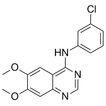
| Description |
AG-1478 is a selective EGFR tyrosine kinase inhibitor with IC50 of 3 nM.
|
| Related Catalog |
|
| Target |
EGFR:3 nM (IC50)
|
| In Vitro |
AG-1478 (AG1478) is irreversible for growth regulation of human lung (A549) and prostate (DU145) cancer cell lines, cultured in chemically defined DMEM/F12 medium. AG-1478 seems to be more effective at lower concentrations, but is unable to completely inhibit growth of A549 cells[1]. Inhibition of EGFR by specific tyrosine kinase inhibitor AG-1478 (AG1478) significantly decreases the angiotensin II-mediated synthesis of TGF-β and fibronectin by cardiac fibroblasts. EGFR is pharmacologically inhibited by small-molecule inhibitor AG-1478 with IC50 of 4 nM[2]. Both Polyfect (PF) and Superfect (SF) treatment lead to increased apoptosis in HEK 293 cells to a similar extent as assessed by flow cytometry. The antioxidant, tempol, significantly reduced dendrimer-mediated apoptosis for both PF and SF. AG-1478 (AG1478), at a 10-fold higher dose (100 μM) than used in signaling studies, is used as a positive control and significantly induced apoptosis in HEK 293 cells[3].
|
| In Vivo |
Administration of AG-1478 (AG1478) significantly reduces myocardial inflammation, fibrosis, apoptosis, and dysfunction in both two obese mouse models. ApoE-/- mice are first fed with HFD for 8 weeks (ApoE-HFD), and then administrated with AG-1478 (10 mg/kg/day) or 542 (10 mg/kg/day) for another 8 weeks by oral gavage. AG-1478 or 542 treatment blocks HFD induced cardiac EGFR phosphorylation in vivo, without affecting the plasma level of low density lipoprotein (LDL) and total triglyceride (TG)[2]. Administration of EGF (10 nM) leads to a robust and reproducible elevation in EGFR phosphorylation that can be blocked by AG-1478 (AG1478), a known inhibitor of EGFR phosphorylation. Increasing doses of Polyfect (PF) result in a significant reduction in EGF-induced EGFR phosphorylation (p<0.05) but this is to a lesser extent than observed with AG1478[3].
|
| Cell Assay |
DU145 (HTB-81) and A549 (CCL-185) cells are seeded on 96-well plates at concentrations of 4×10³ cells/well in MEM (DU145 cells) or DMEM (A549 cells), supplemented with 100 IU/mL penicillin and 100 μg/mL streptomycin in the presence of 10% FBS. Following 24 h of incubation, the culture medium is replaced with serum-free DMEM/F12 (1:1) supplemented with Transferrin (5 mg/mL), sodium selenite (2 ng/mL) and albumin (0.5 mg/mL) [DMEM/F12+]. After an additional 24 h of incubation (Day 0), the medium is replaced by serum-free DMEM/F12+ medium containing tyrosine kinase inhibitors: AG494, AG-1478 respectively in concentration ranges 1-20 μM and 0.1-8 μM. The incubation is continued for the next 24 h at 37°C in a humidified atmosphere. The modified crystal violet staining method (CV) and MTT assay are used to determine the influence of the tyrphostins on the proliferation of target cells. The absorbance is measured using a Tecan multiscan plate recorder[1].
|
| Animal Admin |
Mice[2] 28 C57BL/6 or ApoE-/- mice are randomly divided into four weight-matched groups. 7 mice are fed with standard animal low-fat diet containing 10 kcal.% fat, 20 kcal.% protein and 70 kcal.% carbohydrate serve as a normal control group (Control or ApoE-LF), while the remaining 21 mice are fed with high-fat diet containing 60 kcal.% fat, 20 kcal.% protein and 20 kcal.% carbohydrate for 16 weeks. Since 9th week, AG-1478 or 542 are given daily by oral gavage at a dose of 10 mg/kg/day for the next 8 weeks. Mice in the Control and HFD groups are gavaged with vehicle (1% CMC-Na solution) only. At the day before the sacrifice of ApoE-/- mice, doppler analysis is performed to determine the pathologic cardiac hypertrophy. Rats[3] Male Wistar rats weighing about 300g are used in this study and divided into the following groups (N=5). Group 1: Non-diabetic (Control, C) animals, Group 2: C+PF (10mg/kg administered as a single intraperotoneal (i.p) injection) Group 3: C+SF (10mg/kg i.p); Group 4: C+AG-1478 (1 mg/kg i.p). Group 5: Rats bearing 4 weeks of diabetes (D) induced by a single i.p. injection of streptozotocin (55 mg/kg body weight); Group 6: D+PF (10 mg/kg i.p) Group 7: D+SF (10 mg/kg i.p) and Group 8: D+AG-1478 (1 mg/kg i.p). AG-1478 and dendrimer treatments are administered as single dose for 24h prior to sacrifice. Rat body weight and basal glucose levels are assessed before and after treatments just before sacrificing the animals. An automated blood glucose analyzer is used to assess blood glucose concentrations and rats with a blood glucose concentration above 250 mg/dL (approx. 14 mM) are declared diabetic as in previous studies.
|
| References |
[1]. Bojko A, et al. The effect of tyrphostins AG494 and AG1478 on the autocrine growth regulation of A549 and DU145 cells. Folia Histochem Cytobiol. 2012 Jul 5;50(2):186-95. [2]. Li W, et al. EGFR Inhibition Blocks Palmitic Acid-induced inflammation in cardiomyocytes and Prevents Hyperlipidemia-induced Cardiac Injury in Mice. Sci Rep. 2016 Apr 18;6:24580. [3]. Akhtar S, et al. Cationic Polyamidoamine Dendrimers as Modulators of EGFR Signaling In Vitro and In Vivo. PLoS One. 2015 Jul 13;10(7):e0132215.
|


