| In Vitro |
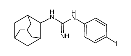
Sigma1 inhibition by IPAG causes the autolysosomal degradation of PD-L1 in PC3 (hormone-insensitive prostate cancer) and MDA-MB-231 (triple-negative breast cancer) cell lines and reduces the levels of functional PD-L1 on the surface of the cells[2]. IPAG treatment produces a mean of 100±8 μg per 106 cells. IPAG can inhibit cell proliferation. Treatment with IPAG decreases cell mass[3]. IPAG treatment suppresses phosphorylation of translational regulator proteins p70S6K, S6, and 4E-BP1[3]. Cell Viability Assay[3] Cell Line: T47D cells Concentration: 10 μM Incubation Time: 24 hours Result: The mean forward scatter height (FSC-H) of DMSO (control) measured 412±5, whereas the mean FSC-H of IPAG treated cells was 390±4. Western Blot Analysis[3] Cell Line: T47D cells Concentration: 10 μM Incubation Time: Result: Decreased levels of phospho-threonine 389-p70S6Kinase (P-S6K), phospho-serine 235/236-ribosomal S6 (P-S6), and phospho-serine 65-4E-BP1 (P-4E-BP1).
|
