CellTracker Blue CMF2HC Dye
Modify Date: 2024-01-24 16:02:03

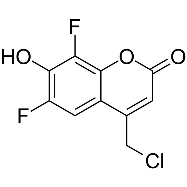
CellTracker Blue CMF2HC Dye structure
|
Common Name | CellTracker Blue CMF2HC Dye | ||
|---|---|---|---|---|
| CAS Number | 215868-45-4 | Molecular Weight | 246.59 | |
| Density | N/A | Boiling Point | N/A | |
| Molecular Formula | C10H5ClF2O3 | Melting Point | N/A | |
| MSDS | N/A | Flash Point | N/A | |
Use of CellTracker Blue CMF2HC DyeCellTracker Blue CMF2HC Dye is a blue dye, can be used in two-channel nuclei acid sequencing, with blue and purple excitation light (450-460 nm/400-405nm or 415-450 nm/480-525nm). CellTracker Blue CMF2HC Dye can be used to rapid determination of antibiotic sensitivity of microorganisms[1][2]. |
| Name | CellTracker Blue CMF2HC Dye |
|---|
| Description | CellTracker Blue CMF2HC Dye is a blue dye, can be used in two-channel nuclei acid sequencing, with blue and purple excitation light (450-460 nm/400-405nm or 415-450 nm/480-525nm). CellTracker Blue CMF2HC Dye can be used to rapid determination of antibiotic sensitivity of microorganisms[1][2]. |
|---|---|
| Related Catalog | |
| In Vitro | CellTracker Blue CMF2HC Dye is a blue dye that can be excitable by a blue light source having a wavelength of about 450-460 nm, is used as the first or the second detectable label described herein[1]. Blue/Violet Two-Channel Sequencing Methods[1]: 1.Contacting a primer polynucleotide/taiget polynucleotide complex with a mixture comprising one or more of a first type of nucleotide, a second type of nucleotide, a third type of nucleotide, and a fourth type of nucleotide, wherein the primer polynucleotide is complementary to at least a portion ofthe single stranded target polynucleotide; 2.Incorporating one type of nucleotide from the mixture to the primer polynucleotide to produce an extended primer polynucleotide (i.e., an extended primer polynucleotide/target polynucleotide complex); 3.Performing a first imaging event using a first excitation light source and collecting a first emission signal from the extended primer polynucleotide/target polynucleotide complex with a first emission filer; 4.Performing a second imaging event using a second excitation light source and collecting a second emission signal from the extended primer polynucleotide/taiget polynucleotide complex with a second emission filter; Note[1]: a. one of the first excitation light source and the second excitation light source has a wavelength of about 350 nm to about 410 nm, and the other one of the first excitation light source and the second excitation light source has a wavelength of about 450 nm to about 460 nm; b. one of the first emission filter and the second emission filter has a detection wavelength of about 415 nm to about 450 nm, and the other one of the first emission filter and the second emission filter has a detection wavelength of about 480 nm to about 525 nm. |
| References |
[2]. Super Michael, et al. Rapid antibiotic susceptibility testing: US, US20150064703[p]. 2015-03-05. |
| Molecular Formula | C10H5ClF2O3 |
|---|---|
| Molecular Weight | 246.59 |