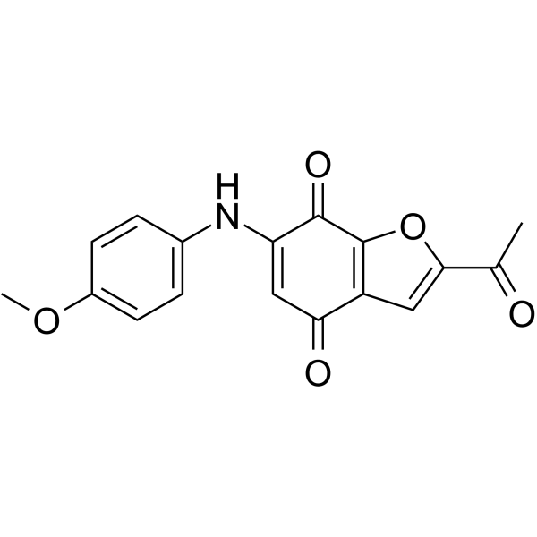
| In Vitro |
STAT3-IN-10 (A11) (48 h) shows IC50 values of 0.67, 0.77, 1.24 µM against MDA-MB-231, MDA-MB-468, HepG2 cells, respectively[1]. STAT3-IN-10 (A11) directly binds to the STAT3 SH2 domain [1]. STAT3-IN-10 (A11) (0-3 μM, 24 h) inhibits the phosphorylation of STAT3 and its downstream target proteins and has a good selectivity against the tumor suppressor STAT1 [1]. STAT3-IN-10 (A11) (0-4 μM, 24 h) induces apoptosis in cancer cells[1]. STAT3-IN-10 (A11) (0-4 μM, 24 h) dose-dependently causes a significant S phase arrest in MDA-MB-231 cells[1]. Cell Viability Assay[1] Cell Line: Breast cancer cell lines: MDA-MB-231 and MDA-MB-468; human liver carcinoma cell line: HepG2. Concentration: Incubation Time: 48 h, re-incubated for 4 h (MDA-MB-468, MDA-MB-231) and 1h (HepG2). Result: Showed IC50 values of 0.67, 0.77, 1.24 µM against MDA-MB-231, MDA-MB-468, HepG2 cells, respectively. Western Blot Analysis[1] Cell Line: MDA-MB-231. Concentration: 0, 0.75, 1.5 and 3.0 μM. Incubation Time: 24 h. Result: Decreased the STAT3-Y705 phosphorylation without affecting the total amount of STAT3 protein and decreased the expression of STAT3 target genes, including C-Myc and Cyclin D1 in a dose-dependent manner. Had little impact on the level of STAT1 and its phosphorylation on Tyr701. Apoptosis Analysis[1] Cell Line: MDA-MB-231. Concentration: 0, 1, 2, and 4 μM. Incubation Time: 24 h. Result: Induced the apoptosis in MDA-MB-231 cells in a concentration-dependent manner Cell Cycle Analysis[1] Cell Line: MDA-MB-231. Concentration: 0, 1, 2, and 4 μM. Incubation Time: 24 h. Result: Could dose-dependently cause a significant S phase arrest in MDA-MB-231 cells
|
