| Description |
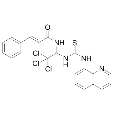
Salubrinal is a cell-permeable and selective inhibitor of eIF2α dephosphorylation.
|
| Related Catalog |
|
| Target |
IC50: 1.7 µM (PP1)[1]
|
| In Vitro |
Salubrinal, a recently identified PP1 inhibitor capable to protect against endoplasmic reticulum (ER) stress in various model systems, strongly synergized with proteasome inhibitors to augment apoptotic death of different leukemic cell lines. Salubrinal preferentially seems to target the PP1/GADD34 complex, Salubrinal is of interest to examine whether the effect of Salubrinal could also be recapitulated by another inhibitor of this phosphatase. For this purpose cantharidin, wis selected, which is less toxic than okadaic acid, but which also blocks PP1 (IC50=1.7 µM) activities[1].
|
| In Vivo |
Salubrinal is a synthetic chemical that inhibits de-phosphorylation of eukaryotic translation initiation factor 2 alpha (eIF2α). Salubrinal significantly suppresses inflammation of the paws of CAIA mice. For instance, the clinical scores are 1.94±1.7 (placebo) and 0.31±0.6 (Salubrinal) on day 6; and 4.63±3.4 (placebo) and 1.09±1.6 (Salubrinal) on day 12. Consistent with the clinical scores, the thickening of the paws is also reduced in the Salubrinal-treated group. Furthermore, Salubrinal reduces the histological scores from 1.47±1.10 (N=16; placebo) to 0.59±0.64 (N=16; Salubrinal) (p=0.01)[2].
|
| Kinase Assay |
Phosphatase activities are determined on immunoprecipitates of the phosphatases. Briefly, 2×10⁶ K562 cells are treated for 18 hr with Salubrinal (20 µM), PSI (10 nM), the combination of both drugs or okadaic acid (100 nM). After washing with PBS, cells are lysed for 15 min on ice either in PP1LB (for determination of PP1γ-activity; 20 mM Tris-HCl, pH 7.5, 1% Triton X-100, 10% glycerol, 132 mM NaCl, Roche complete protease inhibitor ) or in RIPA (for PP2A), supplemented with Roche complete protease inhibitor). Cell lysates containing 500 µg (PP1γ) or 300 µg (PP2A) protein are immunoprecipitated overnight at 4°C with 2-3 µg of the appropriate antibodies and then incubated with Protein A-Sepharose. Immunoprecipitates are washed three times in lysis buffer, followed by resuspension in phosphatase assay buffer (PP2A: 20 mM Tris-HCl, pH7.5, 0.1 mM CaCl2; PP1γ: 50 mM Tris HCl pH 7.0, 0.2 mM MnCl2, 0.1 mM CaCl2, 125 µg/mL BSA, 0.05% Tween 20), supplemented with 100 µM 6,8-difluoro-4-methyl-umbelliferyl phosphate (DiFMUP). Precipitates are allowed to react with substrate for 1 hr at 37°C on an Eppendorf Thermoshaker, centrifuged and DiFMU fluorescence is measured on a BioTek Lambda Fluoro 320 microplate reader (360 nmex/460 nmem). Phosphatase activities are given as percent change relative to the control (DMSO treated cells)[1].
|
| Cell Assay |
Cellular viability is assessed by the WST-1 colorimetric assay. Assays are performed on 96 well plates with 2×10⁴ K562 cells/well in triplicate with Salubrinal concentrations ranging from 5-75 µM (total volume of 200 µL, 18 hrs). Untreated cells served as negative control sample[1].
|
| Animal Admin |
Mice[2] Using Balb/c female mice (~nine weeks old), CAIA is induced by intravenous injection of a 2 mg cocktail of ArthritoMAb antibodies on day 0 followed by intraperitoneal injection of 100 µg LPS on day 3. Mice are randomly divided into a placebo group and a Salubrinal-treated group. Salubrinal (2.0 mg/kg) is intravenously administered daily from day 0, while a solvent (49.5% PEG 400 and 0.5% Tween 80 in PBS) is administered to the placebo group.
|
| References |
[1]. Drexler HC. Synergistic Apoptosis Induction in Leukemic Cells by the Phosphatase Inhibitor Salubrinal and Proteasome Inhibitors. PLoS One. 2009;4(1):e4161. [2]. Hamamura K, et al. Salubrinal acts as a Dusp2 inhibitor and suppresses inflammation in anti-collagen antibody-induced arthritis. Cell Signal. 2015 Apr;27(4):828-35.
|


