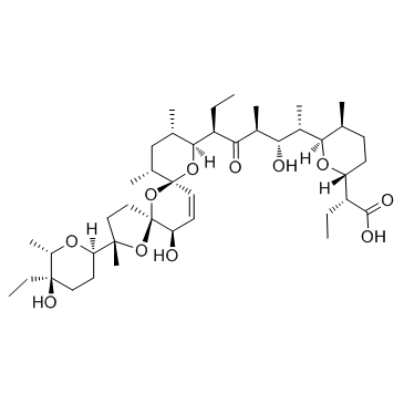
Salinomycin sodium salt
Modify Date: 2025-08-24 15:39:34

Salinomycin sodium salt structure
|
Common Name | Salinomycin sodium salt | ||
|---|---|---|---|---|
| CAS Number | 55721-31-8 | Molecular Weight | 772.98 | |
| Density | N/A | Boiling Point | N/A | |
| Molecular Formula | C42H69NaO11 | Melting Point | 140-142ºC | |
| MSDS | Chinese USA | Flash Point | N/A | |
Use of Salinomycin sodium saltSalinomycin sodium salt is an anticoccidial drug with potent anti-bacterial activity and an novel anticancer agent targeting human cancer stem cells. |
| Name | Salinomycin |
|---|---|
| Synonym | More Synonyms |
| Description | Salinomycin sodium salt is an anticoccidial drug with potent anti-bacterial activity and an novel anticancer agent targeting human cancer stem cells. |
|---|---|
| Related Catalog | |
| Target |
bacterial[1] |
| In Vitro | Salinomycin (0.1-8 µM) inhibits the growth of HUVECs in a dose-dependent manner, accounting for 32.1 and 59.2% inhibition at 4 and 8 µM, respectively. HUVECs exposed to 2, 4 and 8 µM of Salinomycin for 48 h show a dose-dependent reduction in cell number and a change in cell morphology. Salinomycin (4 µM) treatment effectively inhibits HUVEC migration and invasion, and significantly disrupt the capillary-like tube formation of HUVECs. Salinomycin significantly suppresses the expression levels of phosphorylated (p)-FAK in a time- and dose-dependent manner in HUVECs. Salinomycin inhibits HUVEC angiogenesis by disturbing the VEGF-VEGFR2-AKT signaling axis[1]. Combination of RSVL and Salinomycin synergistically inhibits the proliferation of TNBC (MDA-MB-231) cells. RSVL and Salinomycin effectively reduce wound healing, colony and tumorosphere forming capability in TNBC cells. Synergistic combination of RSVL and Salinomycin induces apoptosis in both culture conditions by significant upregulation of Bax with decreased Bcl-2 expression as comparison to untreated and alone drug treatments[2]. Salinomycin (0, 2, 4, 8 and 16 μM) significantly inhibits the proliferation of A2780 and SK-OV-3 cell lines in a dose- and time-dependent manner, (IC50 24h: 13.8 μM, IC50 48h: 6.888 μM and IC50 72h: 4.382 μM for A2780 cell lines), (IC50 24h: 12.7 μM, IC50 48h: 9.869 μM and IC50 72h: 5.022 μM for SK-OV-3 cell lines). Salinomycin blocks the Wnt/β-catenin pathway in EOC cells[3]. Salinomycin (2 μM) reduces cancer cell proliferation, inhibits STAT3 phosphorylation and P38 and β-catenin expressions, and suppresses epithelial-mesenchymal transition in colorectal cancer cells. Salinomycin (1-5 μM) inhibits cancer cell proliferation and STAT3 signaling in colorectal cancer cells. Furthermore, Salinomycin activates Akt (Ser 473) and down-regulates Hsp27 (Ser 82) phosphorylation in HT-29 and SW480. Salinomycin down-regulates hTERT and reduces telomerase activity when combined with telomerase inhibitor[4]. |
| In Vivo | Salinomycin (5 and 10 mg/kg) significantly supresses the average tumor volume and tumor weight. Salinomycin hinders the U251 human glioma cell growth in vivo via inhibition of angiogenesis with involvement of AKT and FAK dephosphorylation[1]. Salinomycin (0.5 mg/kg b.wt.) enhances the mean survival time of the tumor bearing Swiss albino mice[2]. |
| Cell Assay | The effect of Salinomycin on HUVEC growth is determined by MTT assay. Briefly, HUVECs (6,000 cells/well) are seeded in 96-well culture plates for 24 h and incubated with different concentrations of Salinomycin. In the preliminary experiments, Salinomycin treatment for 12, 24, 48 and 72 h shows time-dependent effects on cell growth inhibition. However, treatment for 48 h is the optimal time and is selected for further mechanism evaluation. After Salinomycin treatment for 48 h, 20 µL/well of MTT solution (5 mg/mL) is added and incubated for 5 h. The medium is aspirated and replaced with 200 µL/well of DMSO to dissolve the formazan Salinomycint formed. The color intensity of the formazan solution is measured at 570 nm by a microplate spectrophotometer. The cell viability is expressed as % of the control (as 100%). |
| Animal Admin | Human glioma U251 cells (1×10⁷) suspended in 100 µL PBS are injected into the right lower hind flank of each 6-week-old male nude mouse. The mice are then randomly assigned into three groups of 10 mice in each group. After one week, Salinomycin (5 and 10 mg/kg) is administered into the caudal vein every other day for 16 days. Control mice receive an equal volume of vehicle (Salinomycinine) only. Body weight and tumor volume are monitored every two days. At the end of the experiments, tumors are excised, photographed, and weighed. Tumors from each group are used for western blotting and immunohistochemical (IHC) assay. |
| References |
| Melting Point | 140-142ºC |
|---|---|
| Molecular Formula | C42H69NaO11 |
| Molecular Weight | 772.98 |
| PSA | 173.27000 |
| LogP | 4.78900 |
| Storage condition | 2-8℃ |
| Precursor 0 | |
|---|---|
| DownStream 1 | |
| Salinomycin,monosodium salt |
| Salinomycin hydrate monosodium salt |
| MFCD00167109 |
| salinomycin monosodium salt hydrate |
| salinomycin sodium |
| salinomycin SV sodium salt |
| SALINOMYCIN SODIUM SALT 2.5-HYDRATE |
| SalinoMycin SodiuM Salt ( Min.) |
| Salinomycin 1-sodium salt |
| SalinoMycin HOUSE STANDARD |
| natrium salinomycin |
| Salinomycin (sodium salt) |
 CAS#:53003-10-4