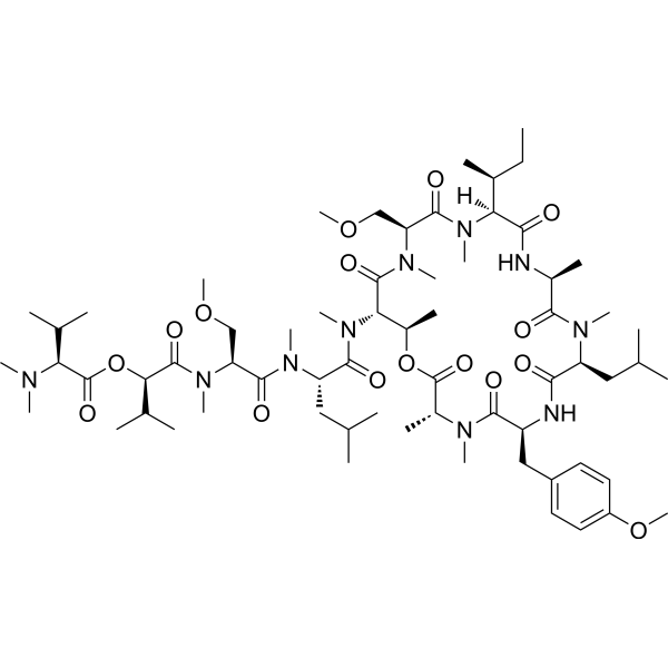
| Description |
Coibamide A, an N-methyl-stabilized cytotoxic depsipeptide, shows potent antiproliferative activity. Coibamide A induces autophagosome accumulation via an mTOR-independent mechanism. Coibamide A induces apoptosis. Coibamide A inhibits VEGFA/VEGFR2 expression and suppresses tumor growth in glioblastoma xenografts[1][2].
|
| Related Catalog |
|
| Target |
VEGFR2
|
| In Vitro |
Coibamide A (0.3-1 nM; 3-60 小时) 抑制 MDA-MB-231 乳腺癌细胞的增殖[1]。 Coibamide A (2.3-230 nM; 3 天) 在人 U87-MG 和 SF-295 胶质母细胞瘤细胞中产生浓度和时间依赖性细胞毒性死亡[2]。 Coibamide A (10-300 nM; 72 小时) 以细胞类型特异性的方式诱导 caspase-3/7 的激活和细胞凋亡[2]。 Coibamide A (20 nM; 48 小时) 在抗凋亡的 U87-MG 细胞中诱导自噬体积累[2]。 Cell Proliferation Assay[1] Cell Line: MDA-MB-231 breast cancer cells Concentration: 0.3, 1 nM Incubation Time: 3-60 hours Result: Showed a steady concentration-dependent decrease in proliferative activity relative to vehicle-treated cells Cell Cytotoxicity Assay[2] Cell Line: U87-MG and SF-295 cells Concentration: 2.3 to 230 nM Incubation Time: 3 days Result: Induced concentration-dependent cytotoxicity with EC50 values of 28.8 nM and 96.2 nM for U87-MG and SF-295 cells, respectively. Apoptosis Analysis[2] Cell Line: U87-MG and SF-295 cells Concentration: 10-300 nM Incubation Time: 72 h Result: An 89 kDa band corresponding to the caspase 3-cleaved form of PARP1 was readily detected by 48 h indicative of apoptotic cell death in SF-295 cells, whereas only trace levels of this fragment were observed in late, detaching U87-MG cell lysates Cell Autophagy Assay[2] Cell Line: U87-MG cell Concentration: 20 nM Incubation Time: 48 h Result: Caused a clear increase in LC3-II expression by 1 h, and this increase in LC3-II expression was generally sustained through 48 h.
|
| In Vivo |
Coibamide A (300 μg/kg; 瘤内注射; 前两天, 之后每 48 小时注射一次, 持续 35 天) 抑制胶质母细胞瘤皮下小鼠模型的肿瘤生长[1]。 Animal Model: 8-week old female nude athymic mice with U87-MG cells[1] Dosage: 300 μg/kg Administration: Intratumoral injections; for the first two days, and then every 48 h afterward for 35 days Result: Remained stable at 200-300 mm3 without significant growth over 4 weeks of treatmen, whereas the tumors of vehicle-treated animals continued to grow at a steady rate consistent with this aggressive cancer cell type
|
| References |
[1]. Jeffrey D Serrill, et al. Coibamide A, a natural lariat depsipeptide, inhibits VEGFA/VEGFR2 expression and suppresses tumor growth in glioblastoma xenografts. Invest New Drugs. 2016 Feb;34(1):24-40. [2]. Andrew M Hau, et al. Coibamide A induces mTOR-independent autophagy and cell death in human glioblastoma cells. PLoS One. 2013 Jun 6;8(6):e65250.
|
