| Structure | Name/CAS No. | Articles |
|---|---|---|
 |
Hydrochloric acid
CAS:7647-01-0 |
|
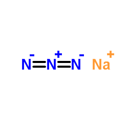 |
Sodium azide
CAS:26628-22-8 |
|
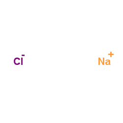 |
sodium chloride
CAS:7647-14-5 |
|
 |
Formaldehyde
CAS:50-00-0 |
|
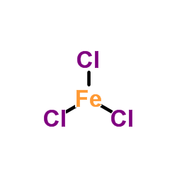 |
Ferric chloride
CAS:7705-08-0 |
|
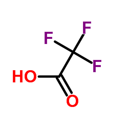 |
trifluoroacetic acid
CAS:76-05-1 |
|
 |
o-xylene
CAS:95-47-6 |
|
 |
Acid Red 87
CAS:17372-87-1 |
|
 |
Eosin Y
CAS:15086-94-9 |
|
 |
SODIUM CHLORIDE-35 CL
CAS:20510-55-8 |