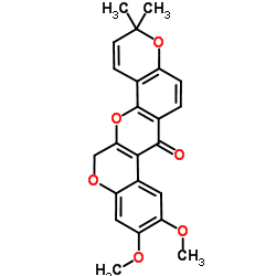
522-17-8
| Name | Deguelin |
|---|---|
| Synonyms |
UNII-K5Z93K66IE
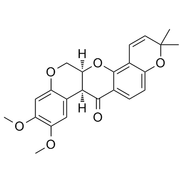
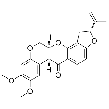
Deguelin Tocris-1770 (7aS,13aS)-9,10-Dimethoxy-3,3-dimethyl-13,13a-dihydro-3H-chromeno[3,4-b]pyrano[2,3-h]chromen-7(7aH)-one MFCD01740600 (-)-cis-Deguelin (7aS-cis)-13,13a-Dihydro-9,10-dimethoxy-3,3-dimethyl-3H-bis[1]benzopyrano[3,4-b:6',5'-e]pyran-7(7aH)-one |
| Description | Deguelin is a natural product isolated from plants in the Mundulea sericea family, and acts as an AKT inhibitor. |
|---|---|
| Related Catalog | |
| Target |
Akt |
| In Vitro | Deguelin (0-500 nM) in a dose and time dependent manner inhibits the growth of MDA-MB-231, MDA-MB-468, BT-549 and BT-20 cells. Deguelin at all concentrations fails to reduce cell numbers in the presence of 1 ng EGF but in the presence of EGF 20 ng reinstated deguelin mediated growth inhibition. Deguelin treated cells show reduced expression of Survivin as determined by western blot and immunofluorescence examinations. Deguelin inhibits p-ERK and its downstream target p-STAT-3 and c-Myc expression in a dose dependent manner[1]. Deguelin down-regulates Akt signaling probably by disrupting its association with Hsp 90 in cultured HNSCC cells. Deguelin deguelin disrupts the association between Hsp 90 with survivin and Cdk4. Deguelin deguelin treatment increases cellular ceramide level through de novo synthase pathway to mediate HNSCC cell death and apoptosis[2]. Deguelin inhibits the proliferation of MPC-11 cells in a concentration- and time-dependent manner and causes the apoptotic death of MPC-11 cells. Following exposure to deguelin, the phosphorylation of Akt is decreased. Deguelin-induced apoptosis is characterized by the upregulation of Bax, downregulation of Bcl-2 and activation of caspase-3[3]. |
| In Vivo | Deguelin (2 or 4 mg/kg, i.p.) reduces the in vivo tumor growth of MDA-MB-231 cells transplanted subcutaneously in athymic mice[1]. Deguelin (4 mg/kg, p.o.) treatment shows a great inhibition in tumor growth, which is demonstrated by reduced tumor size and improved mice survival and, indicating a significant anti-tumor ability by deguelin in vivo[2]. In the colon cancer xenograft model, the volume of the tumor treated with deguelin is significantly lower than that of the control, and the apoptotic index for deguelin-treated mice is much higher[4]. |
| Kinase Assay | Caspase 3 activity is determined using Caspase-Glo-3 assays. This assay provides luminogenic substrate in a buffer system optimized for each specific caspase activity. The caspase cleavage of the substrate is followed by generation of a luminescent signal. The signal generated is proportional to the amount of caspase activity present in the sample. Protein (10 µg) from the cell samples is diluted in water to a final volume of 50 µL and added to a white 96-well microtitre plate, followed by 50 µL of Caspase-Glo-3 reagent. The plate is sealed and gently mixed at 300-500 rpm for 30 s and incubated at room temperature for 30 min. Luminescence is measured in a microplate reader (TECAN Infinite 200). |
| Cell Assay | Breast cancer cells are incubated with increasing concentration of Deguelin ranging from 31 nM to 500 nM for 24, 48 and 72 h. At the termination the cells are trypsinized and cell proliferation is evaluated by counting cells using Z-series Coulter counter. Data are presented as Mean±SE percent of control. |
| Animal Admin | Six to seven weeks old female athymic mice (nu/nu) are housed in a barrier free environment under 24±2°C temperature, 50±10% relative humidity, and 12-hour light/12-hour dark cycle. Mice are provided with sterile mouse chow and water ad libitum. MDA-MB-231 cells (3.0 million cells/animal) are suspended in sterile PBS and then injected subcutaneously into the dorsal flank region using 23 g hypodermic needle. Animals are observed daily for the growth of palpable tumor at the site of injection. Once the tumor (approximately 50 mm3) appears, the mice are randomized in to three groups, animals receiving either 1) vehicle as a control 2) Deguelin treatment at 2 mg/kg bodyweight dose or 3) Deguelin at 4 mg/kg body weight. Each group consists of 10 animals. Vehicle or Deguelin is administered through i.p. injection daily for 21 days. Animals are monitored daily for the signs of drug/vehicle associated toxicity and weighed once weekly. Growth of tumor at the site of cell injection is monitored every alternate day and of tumor size is measured using calipers. Tumor volume is calculated by using the well-established formula: tumor volume (mm3)=π/6 length×width×depth. Data represent the mean tumor volume+SE (mm3) in each group. The animals are sacrificed at the indicated time unless they appear to be moribund or tumors show sign of necrosis. At termination, the tumor is excised, freed from connective tissue and other organs, a small piece is fixed in 10% buffered formalin and remaining tumor is snap frozen for future biochemical analysis. Liver, lung, kidney and spleen are excised and weighed. |
| References |
| Density | 1.3±0.1 g/cm3 |
|---|---|
| Boiling Point | 560.1±50.0 °C at 760 mmHg |
| Melting Point | 85-87ºC(lit.) |
| Molecular Formula | C23H22O6 |
| Molecular Weight | 394.417 |
| Flash Point | 244.7±30.2 °C |
| Exact Mass | 394.141632 |
| PSA | 63.22000 |
| LogP | 5.03 |
| Vapour Pressure | 0.0±1.5 mmHg at 25°C |
| Index of Refraction | 1.584 |
CHEMICAL IDENTIFICATION
HEALTH HAZARD DATAACUTE TOXICITY DATA
|
| Personal Protective Equipment | Eyeshields;Gloves;type N95 (US);type P1 (EN143) respirator filter |
|---|---|
| Safety Phrases | S22 |
| RIDADR | NONH for all modes of transport |
| WGK Germany | 3 |
| RTECS | DX1500000 |
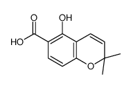
| Precursor 7 | |
|---|---|
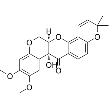
| DownStream 2 | |



![(6aS,12aS)-8-(4-bromo-3-methylbut-2-en-1-yl)-9-hydroxy-2,3-dimethoxy-6,6a-dihydrochromeno[3,4-b]chromen-12(12aH)-one structure](https://image.chemsrc.com/caspic/496/70191-70-7.png)

